Department of Pathology and Laboratory Medicine, Brown University, Providence, RI, USA.
Part Fibre Toxicol. 2011 May 18;8:17. doi: 10.1186/1743-8977-8-17.
The most common causes of granulomatous inflammation are persistent pathogens and poorly-degradable irritating materials. A characteristic pathological reaction to intratracheal instillation, pharyngeal aspiration, or inhalation of carbon nanotubes is formation of epithelioid granulomas accompanied by interstitial fibrosis in the lungs. In the mesothelium, a similar response is induced by high aspect ratio nanomaterials, including asbestos fibers, following intraperitoneal injection. This asbestos-like behaviour of some engineered nanomaterials is a concern for their potential adverse health effects in the lungs and mesothelium. We hypothesize that high aspect ratio nanomaterials will induce epithelioid granulomas in nonadherent macrophages in 3D cultures.
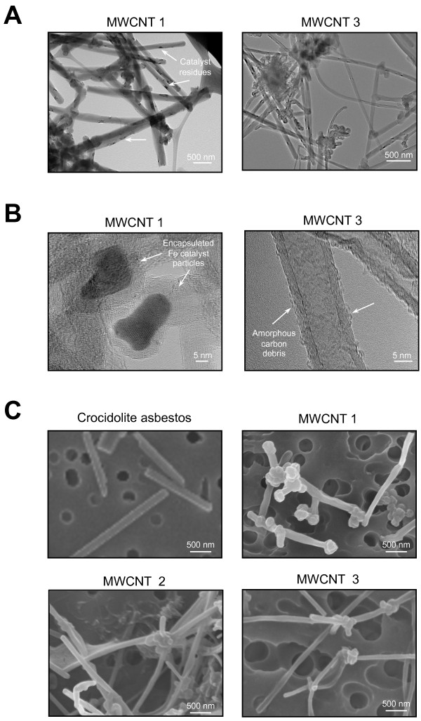
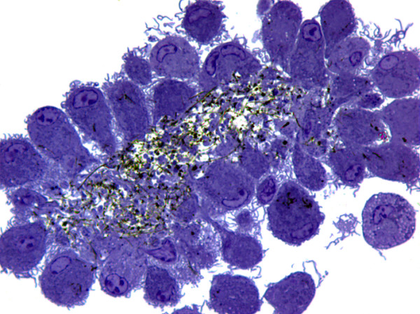
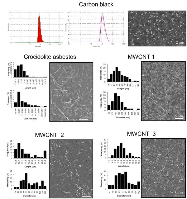
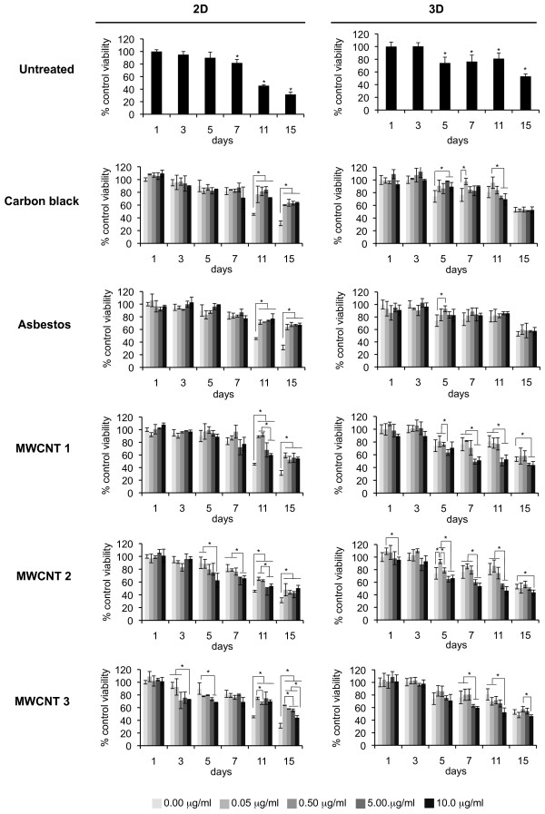
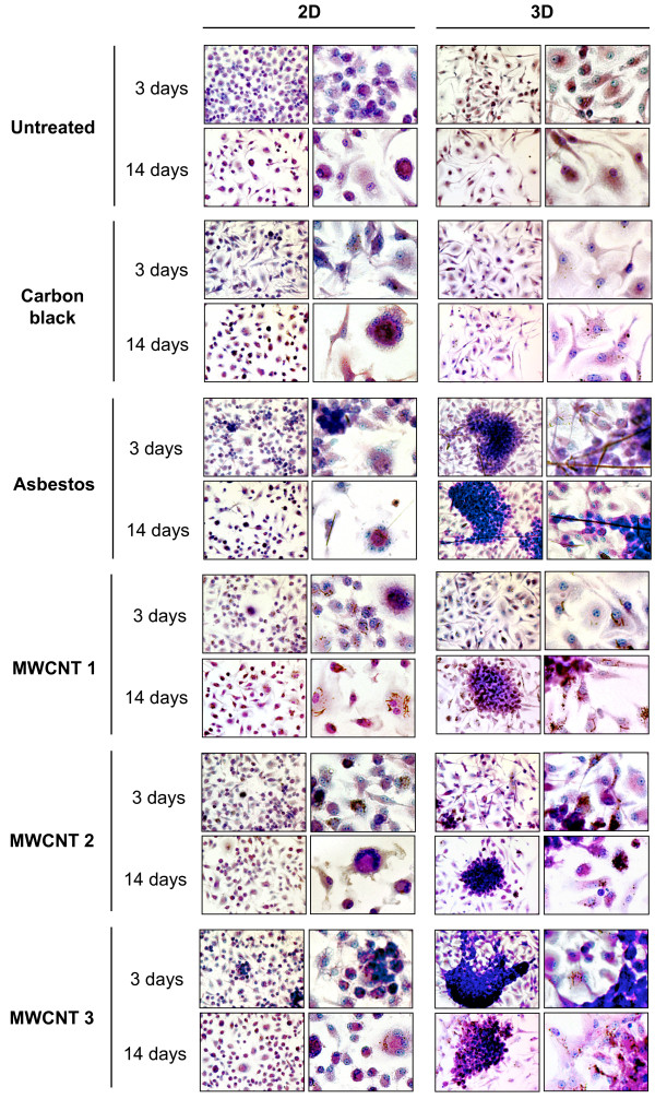
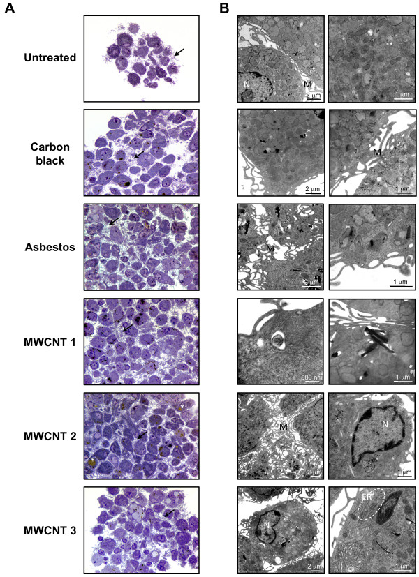
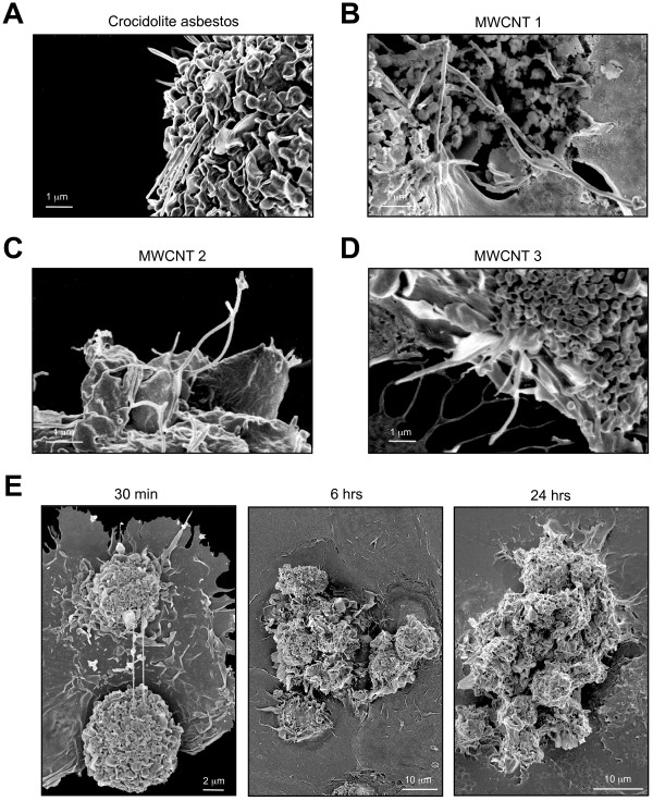
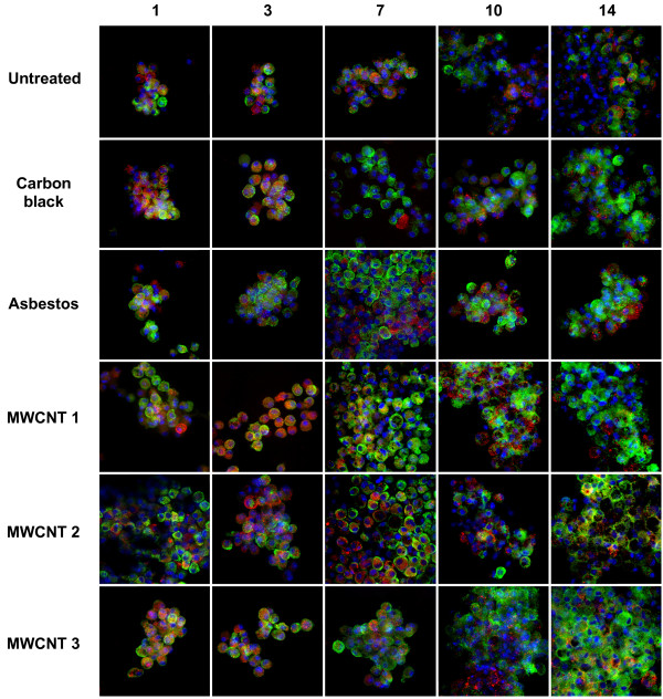
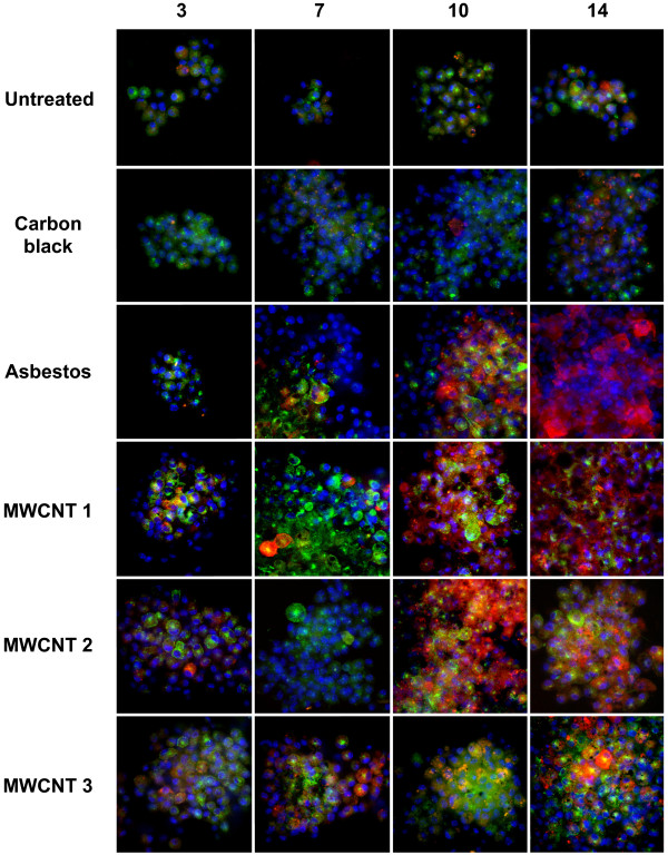
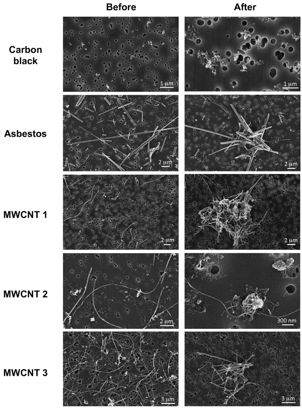
Carbon black particles (Printex 90) and crocidolite asbestos fibers were used as well-characterized reference materials and compared with three commercial samples of multiwalled carbon nanotubes (MWCNTs). Doses were identified in 2D and 3D cultures in order to minimize acute toxicity and to reflect realistic occupational exposures in humans and in previous inhalation studies in rodents. Under serum-free conditions, exposure of nonadherent primary murine bone marrow-derived macrophages to 0.5 μg/ml (0.38 μg/cm2) of crocidolite asbestos fibers or MWCNTs, but not carbon black, induced macrophage differentiation into epithelioid cells and formation of stable aggregates with the characteristic morphology of granulomas. Formation of multinucleated giant cells was also induced by asbestos fibers or MWCNTs in this 3D in vitro model. After 7-14 days, macrophages exposed to high aspect ratio nanomaterials co-expressed proinflammatory (M1) as well as profibrotic (M2) phenotypic markers.
Induction of epithelioid granulomas appears to correlate with high aspect ratio and complex 3D structure of carbon nanotubes, not with their iron content or surface area. This model offers a time- and cost-effective platform to evaluate the potential of engineered high aspect ratio nanomaterials, including carbon nanotubes, nanofibers, nanorods and metallic nanowires, to induce granulomas following inhalation.
引起肉芽肿炎症的最常见原因是持续存在的病原体和难以降解的刺激性物质。将碳纳米管经气管内滴注、咽吸入或吸入肺部后,会出现典型的病理反应,即形成上皮样肉芽肿,并伴有间质纤维化。在间皮中,类似的反应也会由高长径比纳米材料(包括石棉纤维)在腹腔内注射后引起。一些工程纳米材料具有类似石棉的行为,这引起了人们对它们在肺部和间皮中潜在不良健康影响的关注。我们假设高长径比纳米材料将在 3D 培养的非贴壁巨噬细胞中诱导上皮样肉芽肿。
我们使用了炭黑颗粒(Printex 90)和青石棉纤维作为特征明确的参考材料,并将其与三种商业多壁碳纳米管(MWCNT)样品进行了比较。在 2D 和 3D 培养中确定了剂量,以便尽量减少急性毒性,并反映人类实际职业暴露和以前在啮齿动物中的吸入研究。在无血清条件下,将非贴壁原代鼠骨髓来源的巨噬细胞暴露于 0.5μg/ml(0.38μg/cm2)的青石棉纤维或 MWCNT 下,但不暴露于炭黑下,会诱导巨噬细胞分化为上皮样细胞,并形成具有肉芽肿特征形态的稳定聚集体。在这个 3D 体外模型中,MWCNT 或青石棉纤维还诱导多核巨细胞的形成。在 7-14 天后,暴露于高长径比纳米材料的巨噬细胞共同表达了促炎(M1)和促纤维化(M2)表型标志物。
上皮样肉芽肿的形成似乎与碳纳米管的高长径比和复杂 3D 结构相关,而与它们的铁含量或表面积无关。该模型提供了一个时间和成本效益高的平台,可用于评估工程高长径比纳米材料(包括碳纳米管、纳米纤维、纳米棒和金属纳米线)吸入后诱导肉芽肿的潜力。