Department of Pathology and Molecular Medicine, McMaster University, Hamilton, Ontario, Canada.
Diabetes. 2011 Jul;60(7):1964-72. doi: 10.2337/db11-0007. Epub 2011 May 18.
Type 1 diabetes leads to impairments in growth, function, and regenerative capacity of skeletal muscle; however, the underlying mechanisms have not been clearly defined.
With the use of Ins2(WT/C96Y) mice (model of adolescent-onset type 1 diabetes), muscle regeneration was characterized in terms of muscle mass, myofiber size (cross-sectional area), and protein expression. Blood plasma was analyzed for glucose, nonesterified fatty acids, insulin, and plasminogen activator inhibitor-1 (PAI-1). PAI-039, an effective inhibitor of PAI-1, was orally administered to determine if PAI-1 was attenuating muscle regeneration in Ins2(WT/C96Y) mice.
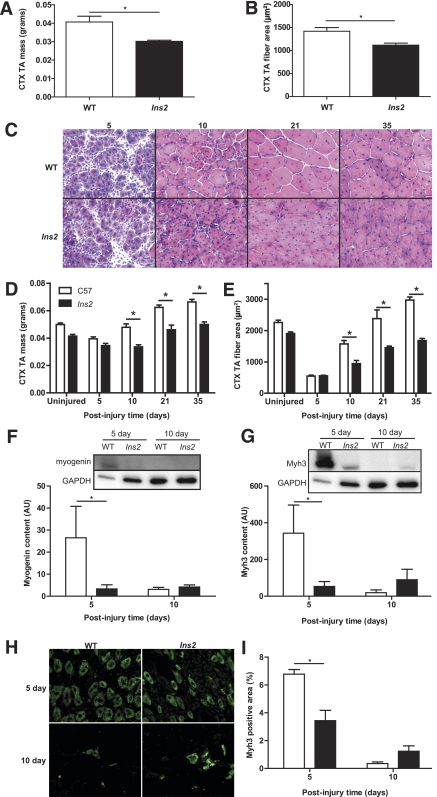
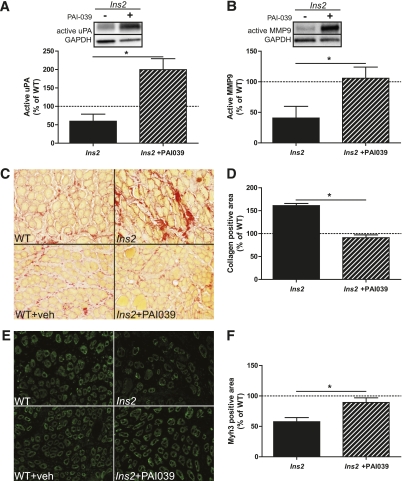
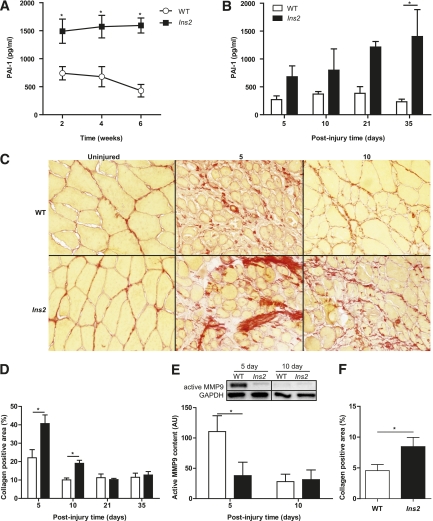
Ins2(WT/C96Y) mice exposed to 1 or 8 weeks of untreated type 1 diabetes before chemically induced muscle injury display significant impairments in their regenerative capacity as demonstrated by decreased muscle mass, myofiber cross-sectional area, myogenin, and Myh3 expression. PAI-1, a physiologic inhibitor of the fibrinolytic system and primary contributor to other diabetes complications, was more than twofold increased within 2 weeks of diabetes onset and remained elevated throughout the experimental period. Consistent with increased circulating PAI-1, regenerating muscles of diabetic mice exhibited excessive collagen levels at 5 and 10 days postinjury with concomitant decreases in active urokinase plasminogen activator and matrix metalloproteinase-9. Pharmacologic inhibition of PAI-1 with orally administered PAI-039 rescued the early regenerative impairments in noninsulin-treated Ins2(WT/C96Y) mice.
Taken together, these data illustrate that the pharmacologic inhibition of elevated PAI-1 restores the early impairments in skeletal muscle repair observed in type 1 diabetes and suggests that early interventional studies targeting PAI-1 may be warranted to ensure optimal growth and repair in adolescent diabetic skeletal muscle.
1 型糖尿病会导致骨骼肌的生长、功能和再生能力受损;然而,其潜在机制尚未明确。
使用 Ins2(WT/C96Y) 小鼠(青少年发病 1 型糖尿病模型),从肌肉质量、肌纤维大小(横截面积)和蛋白表达等方面对肌肉再生进行了特征描述。分析了血浆中的葡萄糖、非酯化脂肪酸、胰岛素和纤溶酶原激活物抑制剂-1(PAI-1)。口服给予 PAI-039,一种有效的 PAI-1 抑制剂,以确定 PAI-1 是否会减弱 Ins2(WT/C96Y) 小鼠的肌肉再生。
在化学诱导肌肉损伤之前,未经治疗的 1 型糖尿病暴露 1 或 8 周的 Ins2(WT/C96Y) 小鼠显示出其再生能力显著受损,表现为肌肉质量、肌纤维横截面积、myogenin 和 Myh3 表达减少。PAI-1 是纤维蛋白溶解系统的生理抑制剂,也是其他糖尿病并发症的主要贡献者,在糖尿病发病后 2 周内增加了两倍以上,并在整个实验期间保持升高。与循环 PAI-1 增加一致,糖尿病小鼠的再生肌肉在损伤后 5 和 10 天表现出过多的胶原水平,同时活性尿激酶纤溶酶原激活物和基质金属蛋白酶-9 减少。口服给予 PAI-039 抑制 PAI-1 可挽救未经胰岛素治疗的 Ins2(WT/C96Y) 小鼠的早期再生损伤。
综上所述,这些数据表明,抑制升高的 PAI-1 可恢复 1 型糖尿病中观察到的骨骼肌修复早期损伤,并表明针对 PAI-1 的早期干预性研究可能是必要的,以确保青少年糖尿病骨骼肌的最佳生长和修复。