Center for Vascular and Inflammatory Diseases, University of Maryland School of Medicine, Baltimore, Maryland 21201, USA.
BMC Immunol. 2011 May 19;12:30. doi: 10.1186/1471-2172-12-30.
Semaphorins were originally identified as molecules regulating a functional activity of axons in the nervous system. Sema4A and Sema4D were the first semaphorins found to be expressed on immune cells and were termed "immune semaphorins". It is known that Sema4A and Sema4D bind Tim-2 and CD72 expressed on leukocytes and PlexinD1 and B1 present on non-immune cells. These neuroimmune semaphorins and their receptors have been shown to play critical roles in many physiological and pathological processes including neuronal development, immune response regulation, cancer, autoimmune, cardiovascular, renal, and infectious diseases. However, the expression and regulation of Sema4A, Sema4D, and their receptors in normal and allergic lungs is undefined.
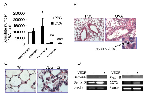
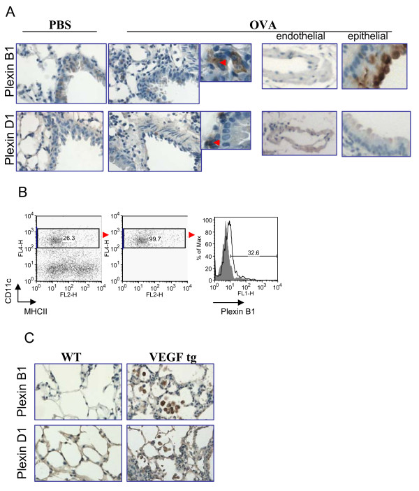
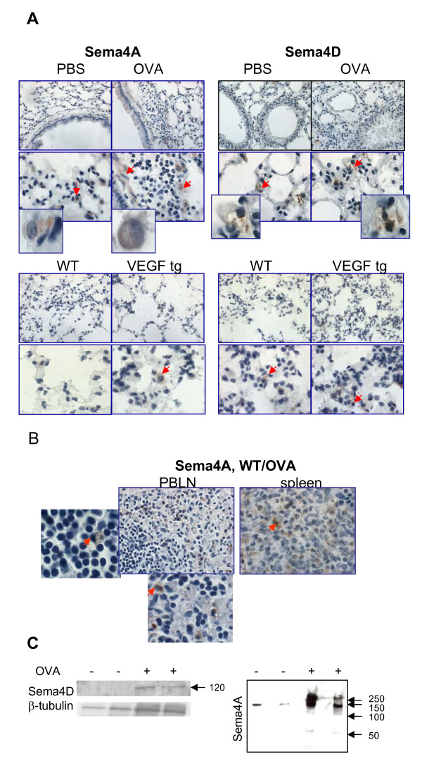
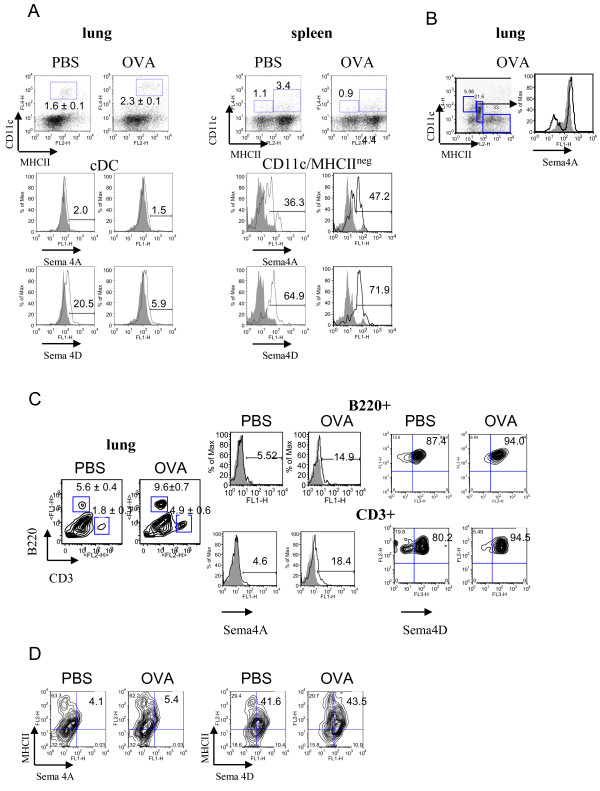
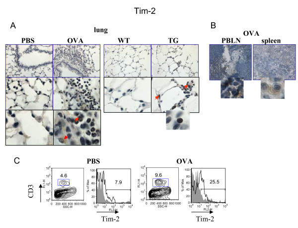
Allergen treatment and lung-specific vascular endothelial growth factor (VEGF) expression induced asthma-like pathologies in the murine lungs. These experimental models of allergic airway inflammation were used for the expression analysis of immune semaphorins and their receptors employing immunohistochemistry and flow cytometry techniques. We found that besides accessory-like cells, Sema4A was also detected on bronchial epithelial and smooth muscle cells, whereas Sema4D expression was high on immune cells such as T and B lymphocytes. Surprisingly, under inflammation various cell types including macrophages, lymphocytes, and granulocytes in the lung expressed Tim-2, a previously defined marker for Th2 cells. CD72 was found on lung immune, inflammatory, and epithelial cells. Bronchial epithelial cells were positive for both plexins, whereas some endothelial cells selectively expressed Plexin D1. Plexin B1 expression was also detected on lung DC. Both allergen and VEGF upregulated the expression of neuroimmune semaphorins and their receptors in the lung tissue. However, the lung tissue Sema4A-Tim2 expression was rather weak, whereas Sema4D-CD72 ligand-receptor pair was vastly upregulated by allergen. Soluble Sema4D protein was present in the lung lysates and a whole Sema4A protein plus its dimer were readily detected in the bronchoalveolar (BAL) fluids under inflammation.
This study clearly shows that neuroimmune semaphorins Sema4A and Sema4D and their receptors might serve as potential markers for the allergic airway inflammatory diseases. Our current findings pave the way for further investigations of the role of immune semaphorins in inflammation and their use as potential therapeutic targets for the inflammatory lung conditions.
信号蛋白最初被鉴定为调节神经系统中轴突功能活动的分子。Sema4A 和 Sema4D 是第一批在免疫细胞中表达的信号蛋白,被称为“免疫信号蛋白”。已知 Sema4A 和 Sema4D 与白细胞上表达的 Tim-2 和 CD72 以及非免疫细胞上的 PlexinD1 和 B1 结合。这些神经免疫信号蛋白及其受体已被证明在许多生理和病理过程中发挥关键作用,包括神经元发育、免疫反应调节、癌症、自身免疫、心血管、肾脏和传染病。然而,Sema4A、Sema4D 及其受体在正常和过敏性肺中的表达和调节尚不清楚。
过敏原处理和肺特异性血管内皮生长因子 (VEGF) 表达在小鼠肺部诱导出哮喘样病理。使用这些过敏性气道炎症的实验模型,采用免疫组织化学和流式细胞术技术分析免疫信号蛋白及其受体的表达。我们发现,除了辅助细胞外,Sema4A 还在支气管上皮细胞和平滑肌细胞上检测到,而 Sema4D 的表达在上皮细胞如 T 和 B 淋巴细胞上较高。令人惊讶的是,在炎症过程中,肺中的各种细胞类型,包括巨噬细胞、淋巴细胞和粒细胞,都表达了 Tim-2,Tim-2 是先前定义的 Th2 细胞标志物。CD72 存在于肺免疫、炎症和上皮细胞上。Plexin 表达于支气管上皮细胞,而一些内皮细胞选择性表达 Plexin D1。肺树突状细胞也表达 Plexin B1。过敏原和 VEGF 均上调了肺组织中神经免疫信号蛋白及其受体的表达。然而,肺组织中 Sema4A-Tim2 的表达较弱,而 Sema4D-CD72 配体-受体对则被过敏原大量上调。可溶性 Sema4D 蛋白存在于肺组织裂解物中,炎症时在支气管肺泡(BAL)液中可轻易检测到完整的 Sema4A 蛋白及其二聚体。
这项研究清楚地表明,神经免疫信号蛋白 Sema4A 和 Sema4D 及其受体可能作为潜在的过敏性气道炎症性疾病的标志物。我们目前的研究结果为进一步研究免疫信号蛋白在炎症中的作用及其作为炎症性肺部疾病的潜在治疗靶点铺平了道路。