Department Cell Biology and Anatomy, University of Miami, Miller School of Medicine, R-124, P.O. Box 016960, Miami, FL 33101, USA.
Virchows Arch. 2011 Sep;459(3):331-8. doi: 10.1007/s00428-011-1102-1. Epub 2011 Jun 12.
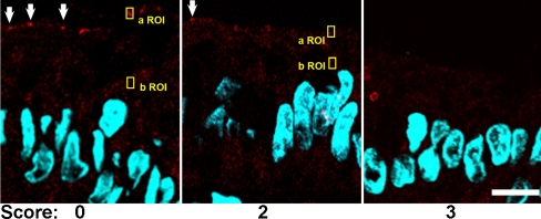
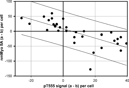
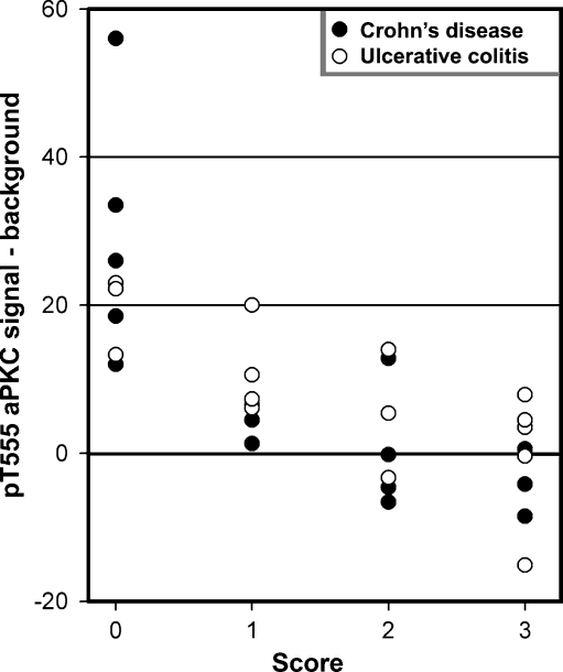
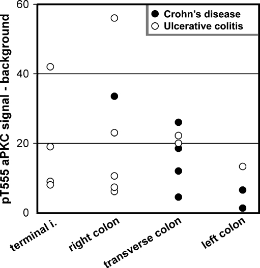
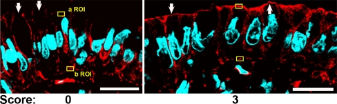
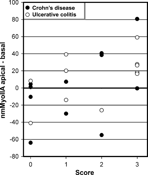
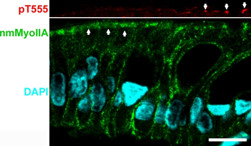
Epithelial barrier function is contingent on appropriate polarization of key protein components. Work in intestinal epithelial cell cultures and animal models of bowel inflammation suggested that atypical PKC (aPKC), the kinase component of the Par3-Par6 polarity complex, is downregulated by pro-inflammatory signaling. Data from other laboratories showed the participation of myosin light chain kinase in intestinal inflammation, but there is paucity of evidence for assembly of its major target, non-muscle myosin II, in inflammatory bowel disease (IBD). In addition, we showed before that non-muscle myosin IIA (nmMyoIIA) is upregulated in intestinal inflammation in mice and TNFα-treated Caco-2 cells. Thus far, it is unknown if a similar phenomena occur in patients with IBD. Moreover, it is unclear whether aPKC downregulation is directly correlated with local mucosal inflammation or occurs in uninvolved areas. Frozen sections from colonoscopy material were stained for immunofluorescence with extensively validated specific antibodies against phosphorylated aPKC turn motif (active form) and nmMyoIIA. Inflammation was scored for the local area from where the material was obtained. We found a significant negative correlation between the expression of active aPKC and local inflammation, and a significant increase in the apical expression of nmMyoIIA in surface colon epithelia in inflamed areas, but not in non-inflamed mucosa even in the same patients. Changes in aPKC and nmMyoIIA expression are likely to participate in the pathogenesis of epithelial barrier function in response to local pro-inflammatory signals. These results provide a rationale for pursuing mechanistic studies on the regulation of these proteins.
上皮屏障功能取决于关键蛋白成分的适当极化。在肠道上皮细胞培养物和肠道炎症的动物模型中的研究表明,非典型蛋白激酶 C(aPKC),即 Par3-Par6 极性复合物的激酶成分,被促炎信号下调。来自其他实验室的数据表明肌球蛋白轻链激酶参与了肠道炎症,但缺乏其主要靶标非肌球蛋白 II(nmMyoII)在炎症性肠病(IBD)中的组装证据。此外,我们之前表明,nmMyoIIA 在小鼠和 TNFα 处理的 Caco-2 细胞中的肠道炎症中上调。到目前为止,尚不清楚 IBD 患者是否会出现类似现象。此外,尚不清楚 aPKC 的下调是否与局部黏膜炎症直接相关,还是发生在未受累区域。使用经过广泛验证的针对磷酸化 aPKC 转角基序(活性形式)和 nmMyoIIA 的特异性抗体对结肠镜检查材料的冷冻切片进行免疫荧光染色。根据获得材料的局部区域对炎症进行评分。我们发现活性 aPKC 的表达与局部炎症之间存在显著的负相关,并且在炎症区域的表面结肠上皮中 nmMyoIIA 的顶端表达显著增加,但在非炎症黏膜中则没有,即使在同一患者中也是如此。aPKC 和 nmMyoIIA 表达的变化可能参与了上皮屏障功能对局部促炎信号的发病机制。这些结果为研究这些蛋白的调节机制提供了依据。