Maisonneuve-Rosemont Hospital Research Center, Montreal, Quebec, Canada.
PLoS One. 2011;6(6):e20705. doi: 10.1371/journal.pone.0020705. Epub 2011 Jun 10.
Epithelial ovarian cancer (EOC) is morphologically heterogeneous being classified as serous, endometrioid, clear cell, or mucinous. Molecular genetic analysis has suggested a role for tumor suppressor genes located at chromosome 3p in serous EOC pathogenesis. Our objective was to evaluate the expression of HYAL1, located at chromosome 3p21.3, in these EOC subtypes, and to investigate its correlation with the expression of steroid hormone receptors.
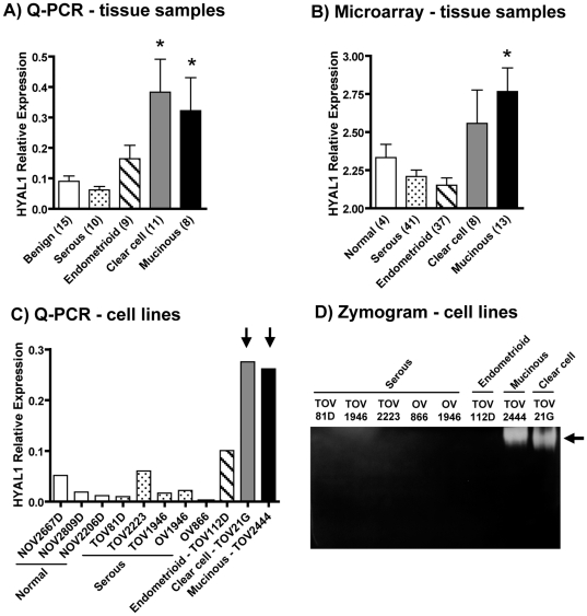
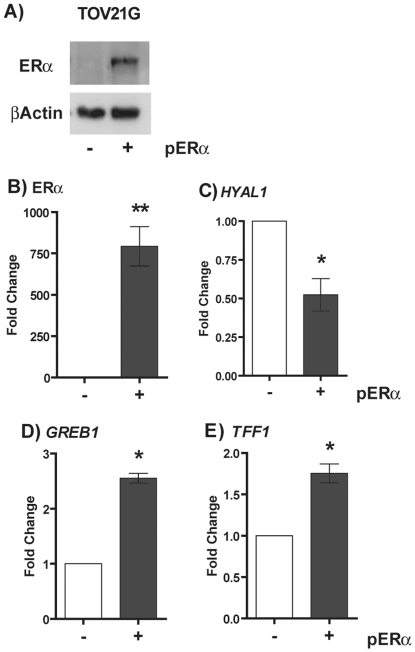
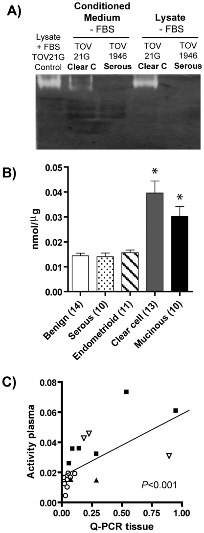
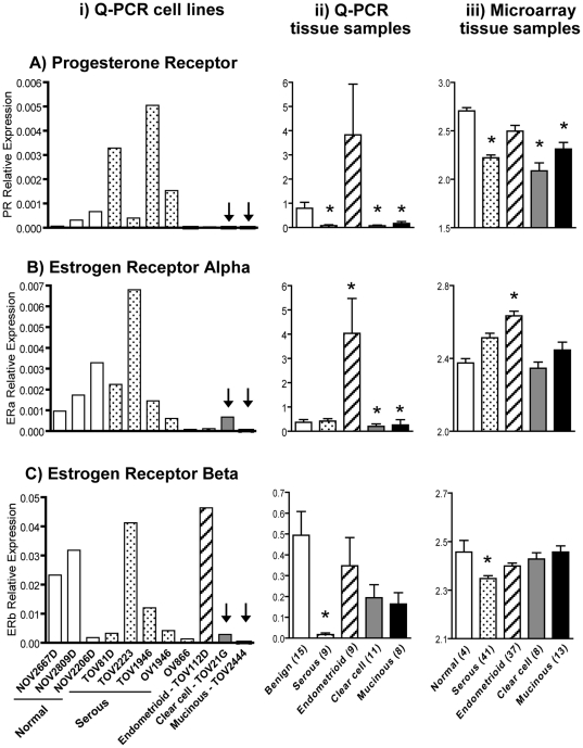
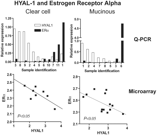
METHODOLOGY/PRINCIPAL FINDINGS: We determined the mRNA expression of HYAL1, estrogen receptor (ER)-α, ERβ and progesterone receptor (PR) in EOC tumor samples and cell lines using quantitative RT-PCR. We also examined the expression of these genes in a publicly available microarray dataset. HYAL-1 enzyme activity was measured in EOC cell lines and in plasma samples from patients. We found that HYAL1 mRNA expression was elevated in clear cell and mucinous EOC tissue samples, but not in serous and endometrioid samples, normal ovaries or benign tumors. Similar results were obtained by two different techniques and with tissue sample cohorts from two independent institutions. Concordantly, HYAL1 mRNA levels and enzymatic activity were elevated only in EOC cell lines derived from clear cell and mucinous subtypes. We also showed that HYAL1 mRNA was inversely correlated to that of ERα specifically in clear cell and mucinous EOCs. Additionally, ectopic expression of ERα in a clear cell EOC cell line (ER- and PR-negative) induced 50% reduction of HYAL1 mRNA expression, supporting a role of ERα in HYAL1 gene regulation. Significantly, HYAL-1 activity was also high in the plasma of patients with these EOC subtypes.
CONCLUSIONS/SIGNIFICANCE: This is the first report showing high HYAL-1 levels in EOC and demonstrating HYAL1 gene repression by ERα. Our results identify Hyaluronidase-1 as a potential target/biomarker for clear cell and mucinous EOCs and especially in tumors with low ERα levels.
上皮性卵巢癌(EOC)在形态上具有异质性,分为浆液性、子宫内膜样、透明细胞或黏液性。分子遗传学分析表明,位于 3p 染色体上的肿瘤抑制基因在浆液性 EOC 的发病机制中起作用。我们的目的是评估位于 3p21.3 上的 HYAL1 在这些 EOC 亚型中的表达,并研究其与甾体激素受体表达的相关性。
方法/主要发现:我们使用定量 RT-PCR 测定 EOC 肿瘤样本和细胞系中 HYAL1、雌激素受体(ER)-α、ERβ 和孕激素受体(PR)的 mRNA 表达。我们还在一个公开的微阵列数据集上检查了这些基因的表达。在 EOC 细胞系和来自患者的血浆样本中测量了 HYAL-1 酶活性。我们发现 HYAL1 mRNA 在透明细胞和黏液性 EOC 组织样本中表达升高,但在浆液性和子宫内膜样样本、正常卵巢或良性肿瘤中不升高。两种不同的技术和来自两个独立机构的组织样本队列得到了相似的结果。一致地,仅在透明细胞和黏液性 EOC 细胞系中,HYAL1 mRNA 水平和酶活性升高。我们还表明,HYAL1 mRNA 与 ERα 呈负相关,特别是在透明细胞和黏液性 EOC 中。此外,在透明细胞 EOC 细胞系(ER-和 PR-阴性)中外源性表达 ERα 可诱导 HYAL1 mRNA 表达降低 50%,支持 ERα 在 HYAL1 基因调控中的作用。重要的是,这些 EOC 亚型患者的血浆中 HYAL-1 活性也很高。
结论/意义:这是首次报道在 EOC 中高表达 HYAL-1,并证明 ERα 抑制 HYAL1 基因。我们的结果将透明细胞和黏液性 EOC 中的 Hyaluronidase-1 确定为潜在的靶点/生物标志物,特别是在 ERα 水平低的肿瘤中。