Department of Physiology, Faculty of Medicine, University of Manitoba, 745 Bannatyne Ave., Winnipeg, MB, Canada R3E 0J9.
Eur J Neurosci. 2011 Jul;34(2):263-71. doi: 10.1111/j.1460-9568.2011.07741.x. Epub 2011 Jun 30.
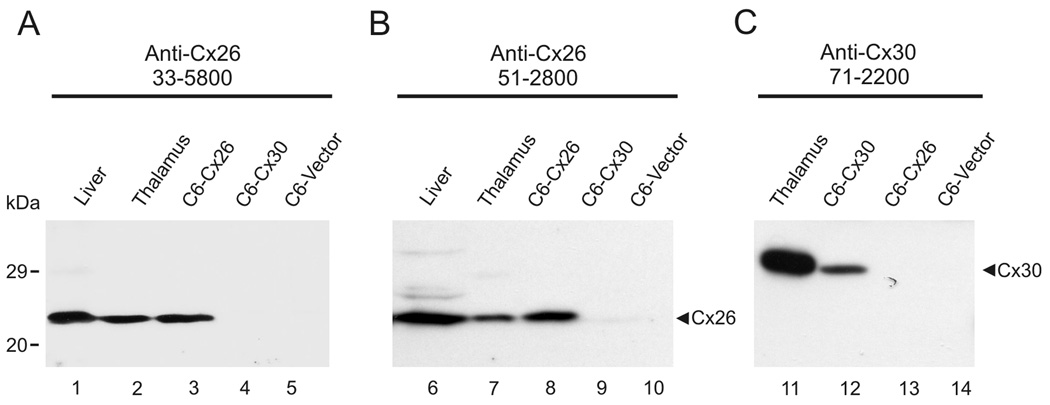
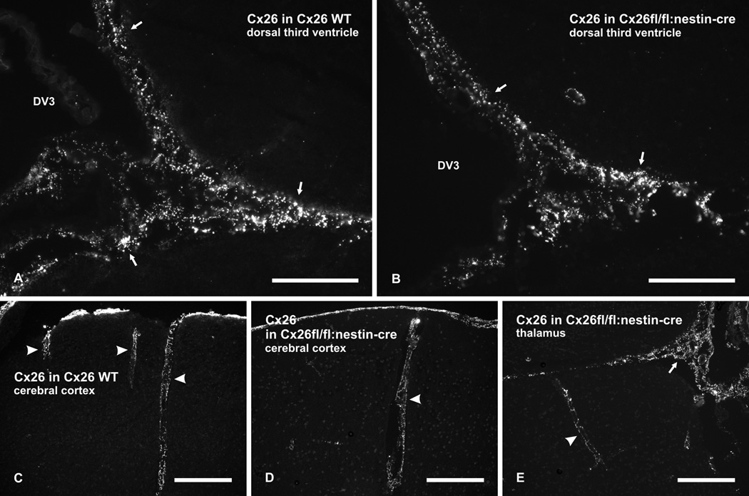
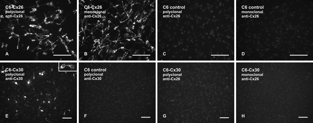
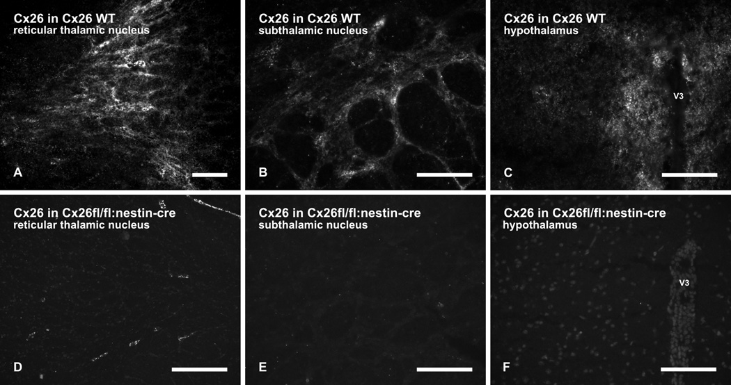
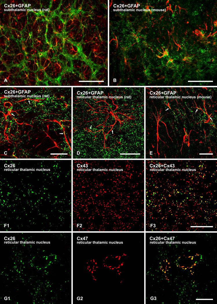
Astrocytes are known to express the gap junction forming proteins connexin30 (Cx30) and connexin43 (Cx43), but it has remained controversial whether these cells also express connexin26 (Cx26). To investigate this issue further, we examined immunofluorescence labelling of glial connexins in wild-type vs. transgenic mice with targeted deletion of Cx26 in neuronal and glial cells (Cx26fl/fl:Nestin-Cre mice). The Cx26 antibodies utilized specifically recognized Cx26 and lacked cross reaction with highly homologous Cx30, as demonstrated by immunoblotting and immunofluorescence in Cx26-transfected and Cx30-transfected C6 glioma cells. Punctate immunolabelling of Cx26 with these antibodies was observed in leptomeninges and subcortical brain regions. This labelling was absent in subcortical areas of Cx26fl/fl:Nestin-Cre mice, but persisted in leptomeningeal tissues of these mice, thereby distinguishing localization of Cx26 between parenchymal and non-parenchymal tissue. In subcortical brain parenchyma, Cx26-positive puncta were often co-localized with astrocytic Cx43, and some were localized along astrocyte cell bodies and processes immunolabelled for glial fibrillary acidic protein. Cx26-positive puncta were also co-localized with punctate labelling of Cx47 around oligodendrocyte somata. Comparisons of Cx26 labelling in rodent species revealed a lower density of Cx26-positive puncta and a more restricted distribution in subcortical regions of mouse compared with rat brain, perhaps partly explaining reported difficulties in detection of Cx26 in mouse brain parenchyma using antibodies or Cx26 gene reporters. These results support our earlier observations of Cx26 expression in astrocytes and its ultrastructural localization in individual gap junction plaques formed between astrocytes as well as in heterotypic gap junctions between astrocytes and oligodendrocytes.
星形胶质细胞被认为表达间隙连接形成蛋白连接蛋白 30(Cx30)和连接蛋白 43(Cx43),但这些细胞是否也表达连接蛋白 26(Cx26)仍存在争议。为了进一步研究这个问题,我们检查了野生型与神经元和神经胶质细胞中 Cx26 靶向缺失的转基因小鼠(Cx26fl/fl:Nestin-Cre 小鼠)中神经胶质连接蛋白的免疫荧光标记。利用这些抗体特异性识别 Cx26,并且缺乏与高度同源的 Cx30 的交叉反应,这一点通过 Cx26 转染和 Cx30 转染 C6 神经胶质瘤细胞的免疫印迹和免疫荧光得到证实。用这些抗体进行的 Cx26 点状免疫标记在软脑膜和皮质下脑区观察到。在 Cx26fl/fl:Nestin-Cre 小鼠的皮质下区域,这种标记缺失,但在这些小鼠的软脑膜组织中仍然存在,从而区分了 Cx26 在实质组织和非实质组织之间的定位。在皮质下脑实质中,Cx26 阳性点状结构通常与星形胶质细胞的 Cx43 共定位,并且一些点状结构定位于星形胶质细胞的细胞体和胶质纤维酸性蛋白免疫标记的突起上。Cx26 阳性点状结构也与少突胶质细胞体周围的 Cx47 点状标记共定位。对不同啮齿动物物种的 Cx26 标记的比较表明,与大鼠脑相比,小鼠脑皮质下区域 Cx26 阳性点状结构的密度较低,分布也较为局限,这可能部分解释了使用抗体或 Cx26 基因报告器在小鼠脑实质中检测到 Cx26 的困难。这些结果支持我们之前的观察结果,即 Cx26 在星形胶质细胞中的表达及其在星形胶质细胞之间形成的单个间隙连接斑和星形胶质细胞与少突胶质细胞之间的异型间隙连接中的超微结构定位。