Faculty of Medical and Human Sciences, University of Manchester, Oxford Road, Manchester M13 9PT, UK.
Eur J Neurosci. 2011 Jul;34(2):213-20. doi: 10.1111/j.1460-9568.2011.07763.x. Epub 2011 Jul 12.
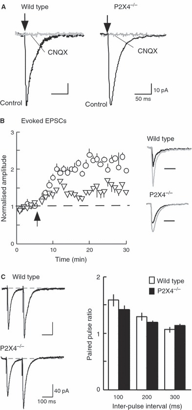
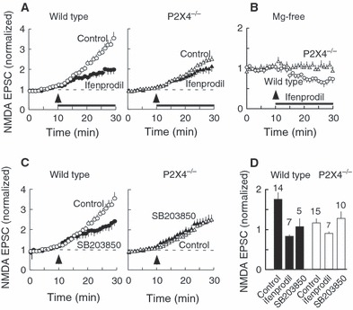
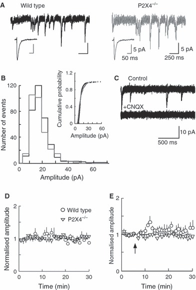
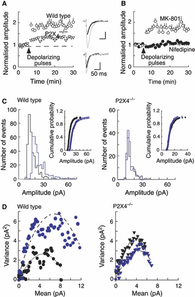
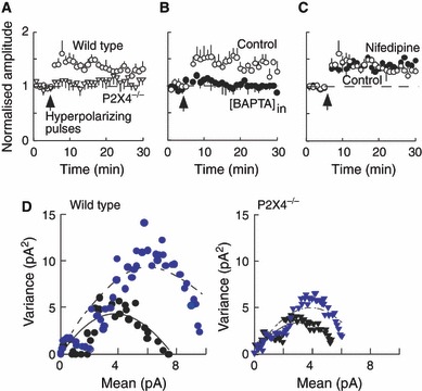
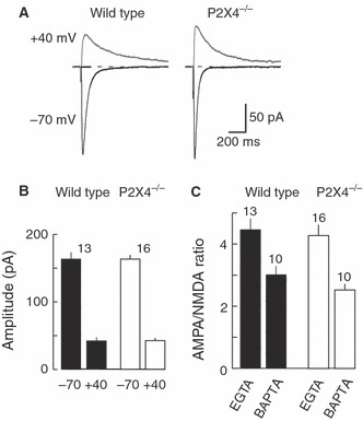
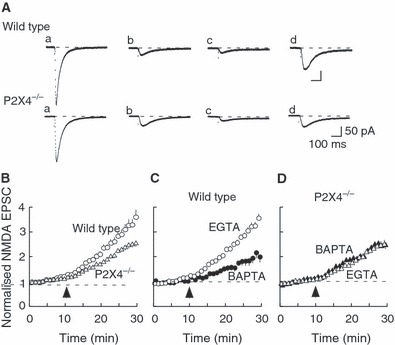
P2X4 receptors are calcium-permeable cation channels gated by extracellular ATP. They are found close to subsynaptic sites on hippocampal CA1 neurons. We compared features of synaptic strengthening between wild-type and P2X4 knockout mice (21-26 days old). Potentiation evoked by a tetanic presynaptic stimulus (100 Hz, 1 s) paired with postsynaptic depolarization was less in P2X4(-/-) mice than in wild-type mice (230 vs. 50% potentiation). Paired-pulse ratios and the amplitude and frequency of spontaneous excitatory postsynaptic currents (EPSCs) were not different between wild-type and knockout mice. Prior hyperpolarization (ten 3 s pulses to -120 mV at 0.17 Hz) potentiated the amplitude of spontaneous EPSCs in wild-type mice, but not in P2X4(-/-) mice; this potentiation was not affected by nifedipine, but was abolished by 10 mM 1,2-bis(o-aminophenoxy)ethane-N,N,N',N'-tetra-acetic acid (BAPTA) in the recording pipette. The amplitude of N-methyl-d-aspartate EPSCs (in 6-cyano-7-nitroquinoxaline-2,3-dione, 10 or 30 μm, at -100 mV) facilitated during 20 min recording in magnesium-free solution. In wild-type mice, this facilitation of the N-methyl-d-aspartate EPSC was reduced by about 50% by intracellular BAPTA (10 mM), ifenprodil (3 μm) or 4-(4-fluorophenyl)-2-(4-methylsulphinylphenyl)-5-(4-pyridyl)1H-imidazole (5 μm). In P2X4(-/-) mice, the facilitation was much less, and was unaffected by intracellular BAPTA, ifenprodil (3 μm) or mitogen-activated protein (MAP) kinase inhibitor 4-(4-fluorophenyl)-2-(4-methylsulphinylphenyl)-5-(4-pyridyl)1H-imidazole (5 μm). This suggests that the absence of P2X4 receptors limits the incorporation of NR2B subunits into synaptic N-methyl-d-aspartate receptors.
P2X4 受体是一种由细胞外 ATP 门控的钙通透性阳离子通道。它们存在于海马 CA1 神经元的突触下部位附近。我们比较了野生型和 P2X4 敲除小鼠(21-26 天龄)之间的突触强化特征。与野生型小鼠相比,由 100 Hz、1 s 的强直刺激引发的突触后去极化诱导的增强作用在 P2X4(-/-)小鼠中较弱(230% vs. 50%增强)。在野生型和敲除型小鼠之间,成对脉冲比、自发兴奋性突触后电流(EPSC)的幅度和频率没有差异。在野生型小鼠中,先前的超极化(10 个 3 s 至 -120 mV 的脉冲,频率为 0.17 Hz)增强了自发 EPSC 的幅度,但在 P2X4(-/-)小鼠中没有;这种增强不受硝苯地平的影响,但在记录电极管中的 10 mM 1,2-双(邻氨基苯氧基)乙烷-N,N,N',N'-四乙酸(BAPTA)中被消除。在无镁溶液中记录 20 分钟期间,N-甲基-D-天冬氨酸 EPSC(在 10 或 30 μm 的 6-氰基-7-硝基喹喔啉-2,3-二酮,-100 mV)的幅度增加。在野生型小鼠中,如果将胞内 BAPTA(10 mM)、ifenprodil(3 μm)或 4-(4-氟苯基)-2-(4-甲基亚磺酰基苯基)-5-(4-吡啶基)1H-咪唑(5 μm)添加到细胞内,则这种 N-甲基-D-天冬氨酸 EPSC 的易化作用会减少约 50%。在 P2X4(-/-)小鼠中,这种易化作用要小得多,并且不受胞内 BAPTA、ifenprodil(3 μm)或丝裂原激活蛋白激酶抑制剂 4-(4-氟苯基)-2-(4-甲基亚磺酰基苯基)-5-(4-吡啶基)1H-咪唑(5 μm)的影响。这表明 P2X4 受体的缺失限制了 NR2B 亚基整合到突触 N-甲基-D-天冬氨酸受体中。