Department of Ultrasound Diagnostics, Tangdu Hospital, Fourth Military Medical University, Xi'an, China.
BMC Gastroenterol. 2011 Jul 19;11:84. doi: 10.1186/1471-230X-11-84.
Abnormality of hepatic vein (HV) waveforms evaluated by Doppler ultrasonography has been widely studied in patients with chronic liver disease. We investigated the correlation between changes in HV waveforms and portal vein velocity (PVVel), the hepatic artery pulsatility index (HAPI), and also the extent of abnormal Doppler HV waveforms expressed as damping index (DI), severity of portal hypertension expressed as Child-Pugh scores and portal pressure (PP) measured directly from patients with portal hypertension (PHT) to evaluate the indicative value of abnormal HV waveforms and discuss the cause of abnormal HV waveform.
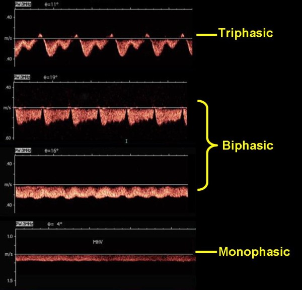
Sixty patients who had been diagnosed with PHT and accepted surgical therapy of portosystemic shunts were investigated. PP was measured intraoperatively. Thirty healthy volunteers with no history of chronic liver disease were enrolled as the control group. HV waveforms were categorized as triphasic, biphasic or monophasic. DI was compared as the quantitative indicator of abnormal HV waveforms. Another two Doppler parameters, PVVel and HAPI were also measured. These Doppler features were compared with PP, Child-Pugh scores and histological changes assessed by liver biopsy.
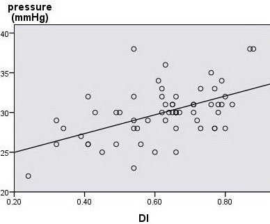
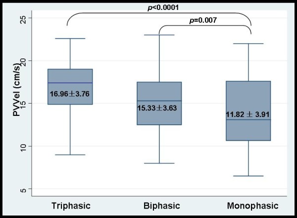
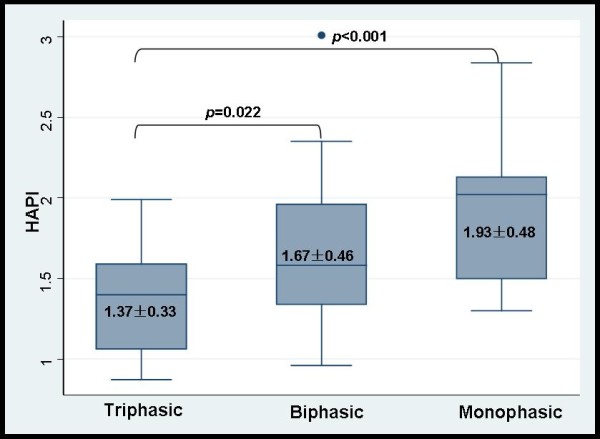
In the patient group, the Doppler flow waveforms in the middle HV were triphasic in 31.6%, biphasic in 46.7%, and monophasic in 21.6% of subjects. These figures were 86.7%, 10.0%, and 3.3%, respectively, in healthy subjects. With the flattening of HV waveforms, the HAPI increased significantly (r = 00.438, p < 0.0001), whereas PVVel decreased significantly (r = -0.44, p <0.0001). Blood flow parameters, HAPI, PVVel and HV-waveform changes showed no significant correlations with Child-Pugh scores. The latter showed a significant correlation with PP (r = 0.589, p = 0.044). Changes of HV waveform and DI significantly correlated with PP (r = 0.579, r = 0.473, p <0.0001), and significant correlation between DI and Child-Pugh scores was observed (r = 0.411, p = 0.001). PP was significantly different with respect to nodule size (p < 0.05), but HV-waveform changes were not significantly correlated with pathological changes.
In patients with PHT, a monophasic HV waveform indicates higher portal pressure. Furthermore, quantitative indicator DI can reflect both higher portal pressure and more severe liver dysfunction. Flattening of HV waveforms accompanied by an increase in the HAPI and decrease in PVVel support the hypothesis that histological changes reducing HV compliance be the cause of abnormality of Doppler HV waveforms from the hemodynamic angle.
通过多普勒超声评估肝静脉(HV)波形异常已在慢性肝病患者中得到广泛研究。我们研究了 HV 波形变化与门静脉速度(PVVel)、肝动脉搏动指数(HAPI)之间的相关性,以及用阻尼指数(DI)表示的异常多普勒 HV 波形的严重程度、用 Child-Pugh 评分表示的门静脉高压严重程度以及直接从门静脉高压(PHT)患者测量的门静脉压力(PP)之间的相关性,以评估异常 HV 波形的指示价值,并探讨异常 HV 波形的原因。
调查了 60 例已被诊断为 PHT 并接受门体分流术治疗的患者。术中测量 PP。选择 30 名无慢性肝病病史的健康志愿者作为对照组。将 HV 波形分为三相、双相或单相。比较 DI 作为异常 HV 波形的定量指标。还测量了另外两个多普勒参数,PVVel 和 HAPI。这些多普勒特征与 PP、Child-Pugh 评分和肝活检评估的组织学变化进行比较。
在患者组中,31.6%、46.7%和 21.6%的中间 HV 多普勒血流波形为三相、双相和单相。在健康受试者中,这些数字分别为 86.7%、10.0%和 3.3%。随着 HV 波形变平,HAPI 显著增加(r=0.438,p<0.0001),而 PVVel 显著降低(r=-0.44,p<0.0001)。血流参数、HAPI、PVVel 和 HV 波形变化与 Child-Pugh 评分无显著相关性。后者与 PP 呈显著相关性(r=0.589,p=0.044)。HV 波形和 DI 的变化与 PP 显著相关(r=0.579,r=0.473,p<0.0001),DI 与 Child-Pugh 评分呈显著相关性(r=0.411,p=0.001)。PP 与结节大小显著相关(p<0.05),但 HV 波形变化与病理变化无显著相关性。
在 PHT 患者中,单相 HV 波形表明门静脉压力较高。此外,定量指标 DI 既能反映较高的门静脉压力,又能反映更严重的肝功能障碍。HV 顺应性降低引起的血流动力学角度异常多普勒 HV 波形的组织学变化假说,HV 波形变平伴 HAPI 增加和 PVVel 降低支持。