Division of Trauma, Surgical Critical Care, and Burns, Department of Surgery, University of California, San Diego School of Medicine, San Diego, CA, USA.
Surgery. 2011 Sep;150(3):379-89. doi: 10.1016/j.surg.2011.06.008. Epub 2011 Jul 23.
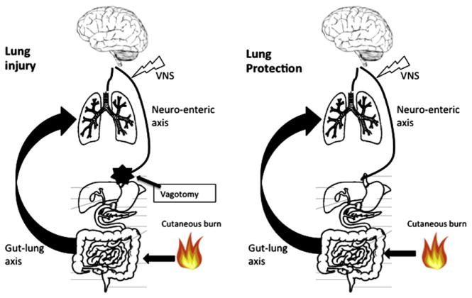
The purpose of this study was to assess acute lung injury when protection to the gut mucosal barrier offered by vagus nerve stimulation is eliminated by an abdominal vagotomy.
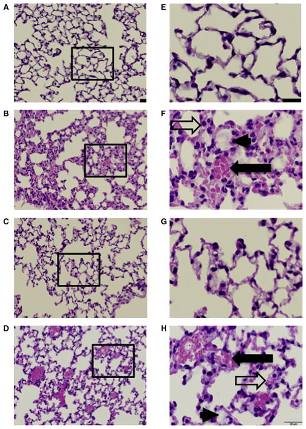
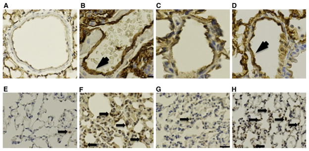
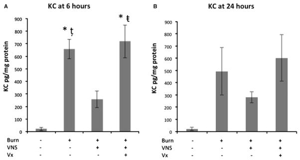
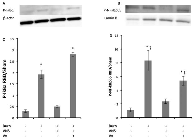
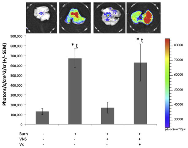
Male balb/c mice were subjected to 30% total body surface area steam burn with and without electrical stimulation to the right cervical vagus nerve. A cohort of animals were subjected to abdominal vagotomy. Lung histology, myeloperoxidase and ICAM-1 immune staining, myeloperoxidase enzymatic assay, and tissue KC levels were analyzed 24 hours after burn. Additionally, lung IkB-α, NF-kB immunoblots, and NF-kB-DNA binding measured by photon emission analysis using NF-kB-luc transgenic mice were performed.
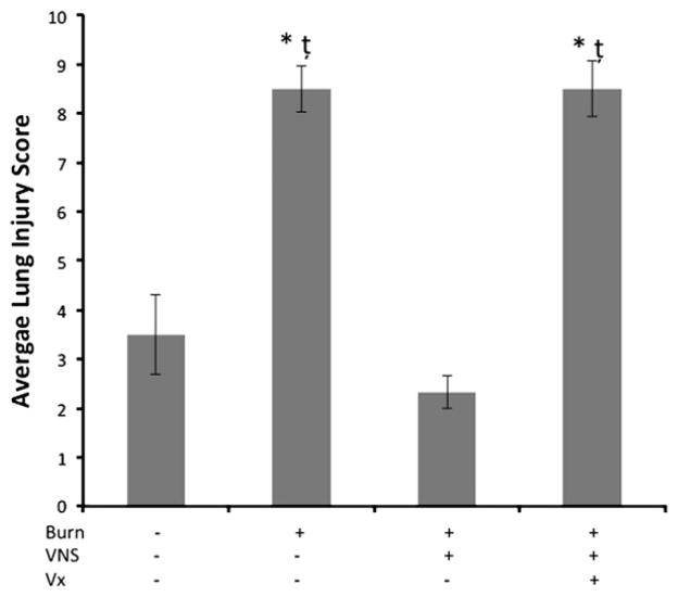
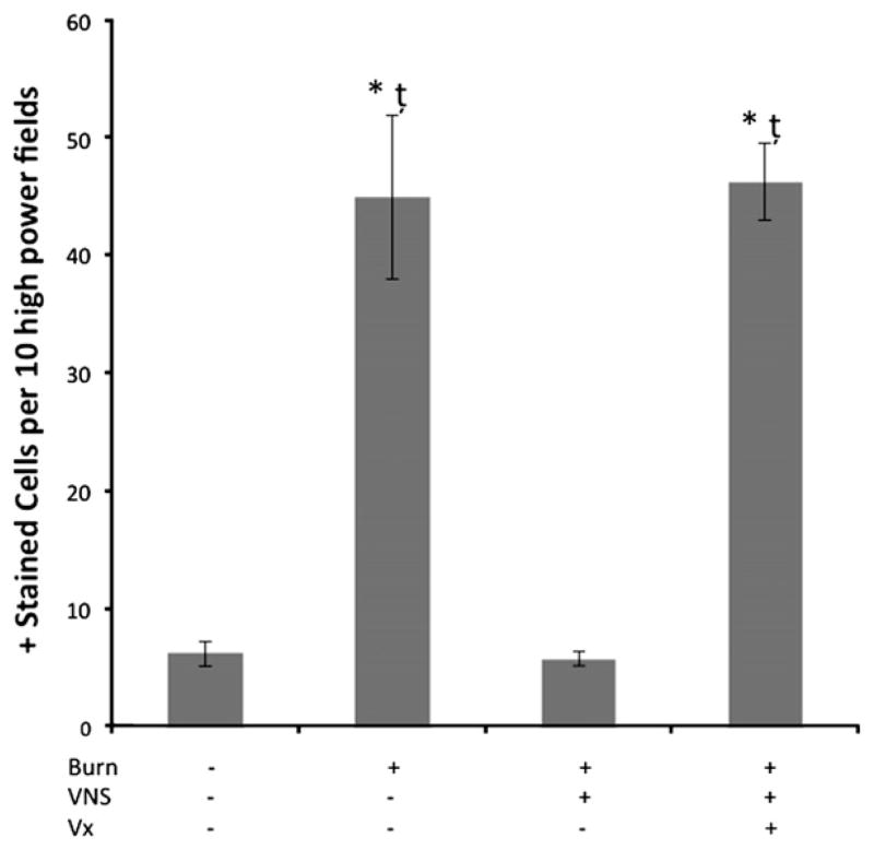
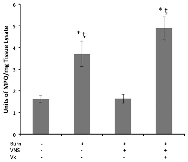
Six hours post burn, phosphorylation of both NF-kB p65 and IkB-α were observed. Increased photon emission signal was seen in the lungs of NF-kB-luc transgenic animals. Vagal nerve stimulation blunted NF-kB activation similar to sham animals whereas abdominal vagotomy eliminated the anti-inflammatory effect. After burn, MPO positive cells and ICAM-1 expression in the lung endothelium was increased, and lung histology demonstrated significant injury at 24 hours. Vagal nerve stimulation markedly decreased neutrophil infiltration as demonstrated by MPO immune staining and enzyme activity. Vagal stimulation also markedly attenuated acute lung injury at 24 hours. The protective effects of vagal nerve stimulation were reversed by performing an abdominal vagotomy.
Vagal nerve stimulation is an effective strategy to protect against acute lung injury following burn. Moreover, the protective effects of vagal nerve stimulation in the prevention of acute lung injury are eliminated by performing an abdominal vagotomy. These results establish the importance of the gut-lung axis after burn in the genesis of acute lung injury.
本研究旨在评估当迷走神经刺激对肠道黏膜屏障的保护作用被腹部迷走神经切断术消除时,急性肺损伤的情况。
雄性 balb/c 小鼠接受 30%的全身表面积蒸汽烧伤,并接受右侧颈迷走神经的电刺激和不接受电刺激。一组动物接受腹部迷走神经切断术。烧伤后 24 小时分析肺组织学、髓过氧化物酶和 ICAM-1 免疫染色、髓过氧化物酶酶活性测定以及组织 KC 水平。此外,还使用 NF-kB-luc 转基因小鼠进行肺 IkB-α、NF-kB 免疫印迹和 NF-kB-DNA 结合的光子发射分析。
烧伤后 6 小时,观察到 NF-kB p65 和 IkB-α的磷酸化。NF-kB-luc 转基因动物的肺部可见增加的光子发射信号。迷走神经刺激类似于假手术动物一样减弱了 NF-kB 的激活,而腹部迷走神经切断术消除了抗炎作用。烧伤后,肺内皮细胞中 MPO 阳性细胞和 ICAM-1 表达增加,肺组织学显示 24 小时后出现明显损伤。迷走神经刺激通过 MPO 免疫染色和酶活性明显减少中性粒细胞浸润。迷走神经刺激也显著减轻了烧伤后 24 小时的急性肺损伤。腹部迷走神经切断术逆转了迷走神经刺激的保护作用。
迷走神经刺激是预防烧伤后急性肺损伤的有效策略。此外,腹部迷走神经切断术消除了迷走神经刺激在预防急性肺损伤中的保护作用。这些结果确立了烧伤后肠-肺轴在急性肺损伤发病机制中的重要性。