Department of Oral Anatomy and Cell Biology School of Dentistry, Pusan National University, Medical Research Institute, Pusan National University Hospital, Yangsan, Korea.
Yonsei Med J. 2011 Sep;52(5):773-8. doi: 10.3349/ymj.2011.52.5.773.
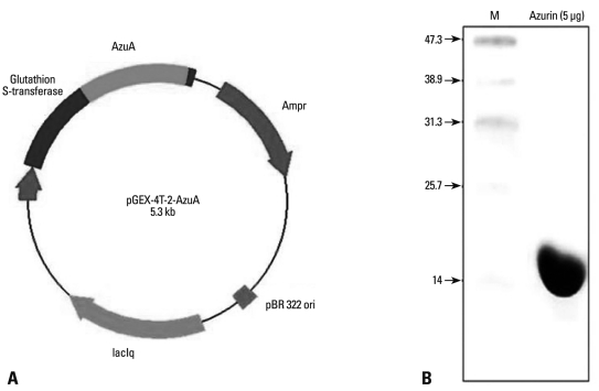
Surgical therapy is the primary treatment for oral cancer, but it can cause facial distortion. Therefore, if anticancer drugs are effective against oral cancer, they may be used preferentially. However, oral squamous carcinoma cells (OSCCs) are resistant to these drugs, so finding a way to enhance the sensitivity of these cells to anticancer drugs is important. The bacterial protein azurin is known to selectively enter cancer cells and induce apoptosis. In this study, we show the anticancer effect of azurin in OSCC.
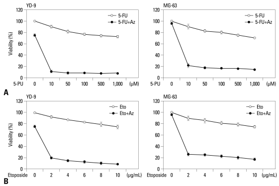
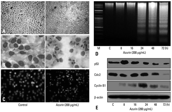
OSCC cell line (YD-9) was subjected to azurin treatment. Cell viability, morphology and protein expression levels were monitored after treatment of azurin. Cells were also subjected to combination treatment of azurin with either 5-fluorouracil or etopside.
Azurin-treated cells showed decreased cell viability accompanied by apoptotic phenotypes including morphological change, DNA breakage, and increases in p53 and cyclin B1 protein levels. Combination treatment of azurin with other anti-tumor agents caused an increase in sensitivity to anticancer drugs in azurin-treated YD-9 cells.
Azurin has a strong synergistic anticancer effect on oral cancer cells when it is used along with anticancer drugs.
手术治疗是口腔癌的主要治疗方法,但会导致面部畸形。因此,如果抗癌药物对口腔癌有效,可能会优先使用。然而,口腔鳞状细胞癌(OSCC)对这些药物有抗药性,因此寻找一种方法来提高这些细胞对抗癌药物的敏感性很重要。已知细菌蛋白蓝铜蛋白选择性进入癌细胞并诱导细胞凋亡。在这项研究中,我们展示了蓝铜蛋白对 OSCC 的抗癌作用。
OSCC 细胞系(YD-9)接受蓝铜蛋白处理。处理蓝铜蛋白后,监测细胞活力、形态和蛋白表达水平。还对蓝铜蛋白与 5-氟尿嘧啶或依托泊苷联合治疗的细胞进行了处理。
用蓝铜蛋白处理的细胞表现出细胞活力降低,同时伴有凋亡表型,包括形态变化、DNA 断裂以及 p53 和细胞周期蛋白 B1 蛋白水平的升高。蓝铜蛋白与其他抗肿瘤药物联合治疗可增加蓝铜蛋白处理的 YD-9 细胞对抗癌药物的敏感性。
当与抗癌药物一起使用时,蓝铜蛋白对口腔癌细胞具有很强的协同抗癌作用。