Department of Radiology and Research Institute of Radiological Science, Yonsei University College of Medicine, Seoul, Korea.
Yonsei Med J. 2011 Sep;52(5):838-44. doi: 10.3349/ymj.2011.52.5.838.
To compare the cytological results of ultrasound-guided fine-needle aspiration (US-FNA) cytology of thyroid nodules to sonographic findings and determine whether US findings are helpful in the interpretation of cytological results.
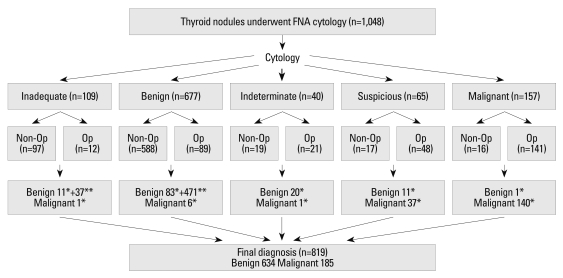
Among the thyroid nodules that underwent US-FNA cytology, we included the 819 nodules which had a conclusive diagnosis. Final diagnosis was based on pathology from surgery, repeated FNA cytology or follow-up of more than one year. Cytological results were divided into five groups: benign, indeterminate (follicular or Hurthle cell neoplasm), suspicious for malignancy, malignant, and inadequate. US findings were categorized as benign or suspicious. Cytological results and US categories were analyzed.
Final diagnosis was concluded upon in 819 nodules based on pathology (n=311), repeated FNA cytology (n=204) and follow-up (n=304), of which 634 were benign and 185 were malignant. There were 560 benign nodules, 141 malignant nodules, 49 nodules with inadequate results, 21 with indeterminate results, and 48 that were suspicious for malignancy. The positive and negative predictive values of the US categories were 59.1% and 97.0%, and those of the cytological results were 93.7% and 98.9%. The US categories were significantly correlated with final diagnosis in the benign (p=0.014) and suspicious for malignancy (p<0.001) cytological result groups, but not in the inadequate and indeterminate cytological results groups. The false positive and negative rates of cytological results were 1.9% and 3.2%.
Sonographic findings can be useful when used alongside cytological results, especially in nodules with cytological results that are benign or suspicious for malignancy.
比较超声引导下甲状腺结节细针抽吸细胞学检查(US-FNA 细胞学)的细胞学结果与超声表现,并确定超声表现是否有助于解释细胞学结果。
在接受 US-FNA 细胞学检查的甲状腺结节中,我们纳入了有明确诊断的 819 个结节。最终诊断基于手术病理、重复 FNA 细胞学或超过 1 年的随访。细胞学结果分为 5 组:良性、不确定(滤泡或 Hurthle 细胞肿瘤)、疑似恶性、恶性和不满意。将超声表现分为良性或可疑。分析细胞学结果和 US 分类。
根据病理(n=311)、重复 FNA 细胞学(n=204)和随访(n=304),对 819 个结节进行了最终诊断,其中 634 个为良性,185 个为恶性。良性结节 560 个,恶性结节 141 个,结果不满意 49 个,不确定 21 个,可疑恶性 48 个。US 分类的阳性和阴性预测值分别为 59.1%和 97.0%,细胞学结果分别为 93.7%和 98.9%。US 分类与良性(p=0.014)和可疑恶性(p<0.001)细胞学结果组的最终诊断显著相关,但与不满意和不确定的细胞学结果组不相关。细胞学结果的假阳性和假阴性率分别为 1.9%和 3.2%。
当与细胞学结果结合使用时,超声表现可能有用,尤其是在具有良性或可疑恶性细胞学结果的结节中。