Department of Translational Medecine and Neurogenetics, IGBMC (Institut de Génétique et de Biologie Moléculaire et Cellulaire), 1 rue Laurent Fries, 67404 Illkirch, France.
Skelet Muscle. 2011 Jul 13;1(1):26. doi: 10.1186/2044-5040-1-26.
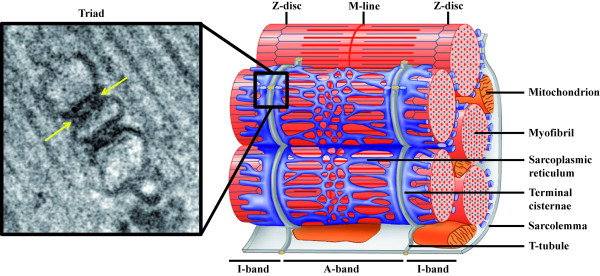
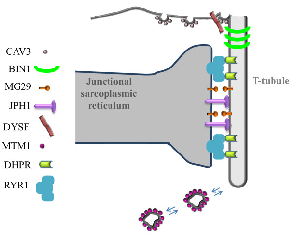
In skeletal muscle, the excitation-contraction (EC) coupling machinery mediates the translation of the action potential transmitted by the nerve into intracellular calcium release and muscle contraction. EC coupling requires a highly specialized membranous structure, the triad, composed of a central T-tubule surrounded by two terminal cisternae from the sarcoplasmic reticulum. While several proteins located on these structures have been identified, mechanisms governing T-tubule biogenesis and triad formation remain largely unknown. Here, we provide a description of triad structure and plasticity and review the role of proteins that have been linked to T-tubule biogenesis and triad formation and/or maintenance specifically in skeletal muscle: caveolin 3, amphiphysin 2, dysferlin, mitsugumins, junctophilins, myotubularin, ryanodine receptor, and dihydhropyridine Receptor. The importance of these proteins in triad biogenesis and subsequently in muscle contraction is sustained by studies on animal models and by the direct implication of most of these proteins in human myopathies.
在骨骼肌中,兴奋-收缩(EC)偶联机制将神经传递的动作电位转化为细胞内钙释放和肌肉收缩。EC 偶联需要高度特化的膜结构——三联体,由中央 T 管周围的两个终池组成,终池来自肌浆网。虽然已经鉴定出位于这些结构上的几种蛋白质,但调节 T 管发生和三联体形成的机制在很大程度上仍然未知。在这里,我们提供了三联体结构和可塑性的描述,并回顾了与 T 管发生和三联体形成和/或维持特别相关的蛋白质在骨骼肌中的作用: caveolin 3、amphiphysin 2、dysferlin、mitsugumins、junctophilins、myotubularin、ryanodine 受体和二氢吡啶受体。这些蛋白质在三联体发生中的重要性,以及随后在肌肉收缩中的重要性,得到了动物模型研究的支持,并且这些蛋白质中的大多数都直接涉及人类肌病。