Psychiatry Neuroimaging Laboratory, Department of Psychiatry, Brigham and Women's Hospital, Harvard Medical School, Boston, MA, USA.
Schizophr Res. 2011 Oct;132(1):69-74. doi: 10.1016/j.schres.2011.07.010. Epub 2011 Aug 9.
Structural abnormalities in the callosal fibers connecting the heteromodal association areas of the prefrontal and temporoparietal cortices bilaterally have been suggested to play a role in the etiology of schizophrenia.
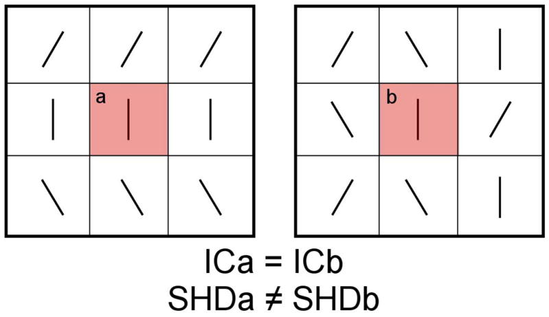
To investigate for geometric abnormalities in these callosal fibers in schizophrenia patients by using a novel Diffusion-Tensor Imaging (DTI) metric of fiber geometry named Shape-Normalized Dispersion (SHD).
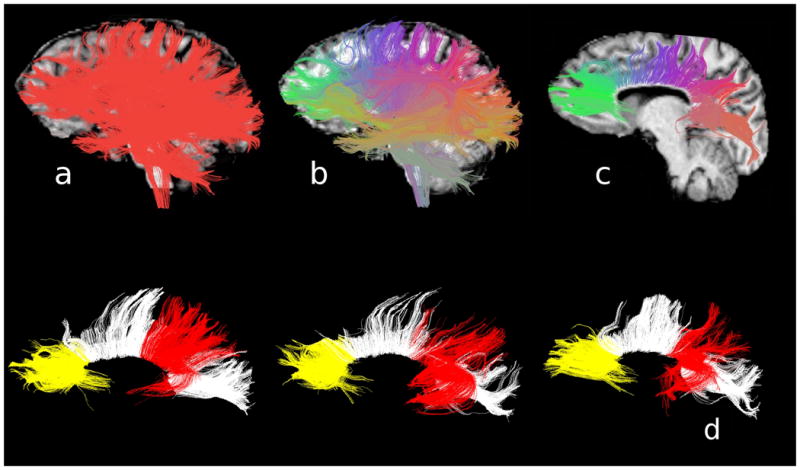
DTIs (3T, 51 gradient directions, 1.7mm isotropic voxels) were acquired from 26 schizophrenia patients and 23 matched healthy controls. The prefrontal and temporoparietal fibers of the corpus callosum were extracted by means of whole-brain tractography, and their mean SHD calculated.
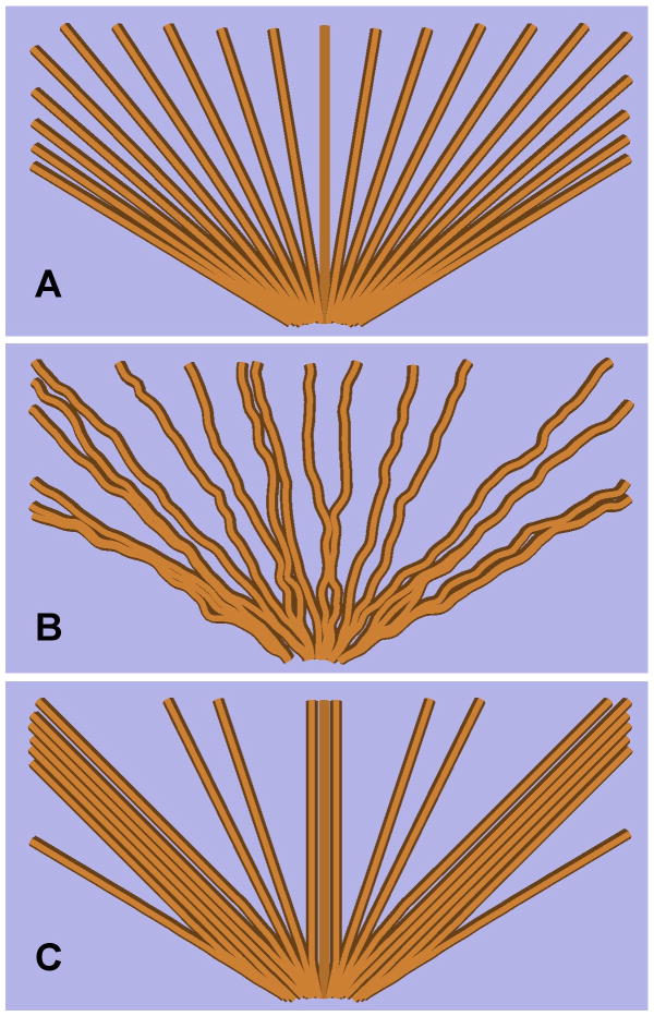
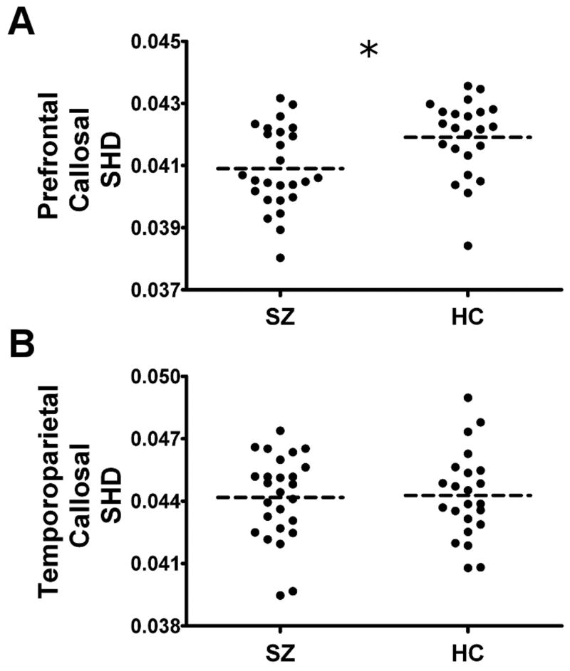
The schizophrenia patients exhibited subnormal levels of SHD in the prefrontal callosal fibers when controlling for between-group differences in Fractional Anisotropy. Reduced SHD could reflect either irregularly turbulent or inhomogeneously distributed fiber trajectories in the corpus callosum.
The results suggest that the transcallosal misconnectivity thought to be associated with schizophrenia could reflect abnormalities in fiber geometry. These abnormalities in fiber geometry could potentially be underpinned by neurodevelopmental irregularities.
双侧额颞叶皮质异模态联合区的胼胝体纤维的结构异常被认为在精神分裂症的病因学中起作用。
通过使用一种新的称为形状归一化离散度(SHD)的纤维几何扩散张量成像(DTI)度量方法,研究精神分裂症患者这些胼胝体纤维的几何异常。
对 26 名精神分裂症患者和 23 名匹配的健康对照组进行了 DTI(3T,51 个梯度方向,1.7mm 各向同性体素)采集。通过全脑追踪技术提取胼胝体的额颞叶纤维,并计算其平均 SHD。
在控制组间各向异性分数差异的情况下,精神分裂症患者的额状纤维 SHD 值低于正常水平。降低的 SHD 可能反映了胼胝体中纤维轨迹的不规则紊乱或不均匀分布。
研究结果表明,与精神分裂症相关的胼胝体间连接异常可能反映了纤维几何形状的异常。这些纤维几何形状的异常可能潜在地受到神经发育不规则的影响。