Liu Gaoqin, Lu Peirong, Li Longbiao, Jin Hui, He Xuefei, Mukaida Naofumi, Zhang Xueguang
Department of Ophthalmology, the First Affiliated Hospital of Soochow University, Suzhou, P.R. China.
Mol Vis. 2011;17:2129-38. Epub 2011 Aug 10.
To address the roles of the stromal derived factor-1 (SDF-1) α in the course of experimental corneal neovascularization (CNV).
CNV was induced by alkali injury and compared in SDF-1α- or vehicle-treated mice two weeks after injury. Angiogenic factor expression in the early phase after injury was quantified by reverse transcription polymerase chain reaction (RT-PCR). Progenitor cell, macrophage, and monocyte intracorneal accumulation in the early phase after injury was evaluated by flow cytometric analysis.
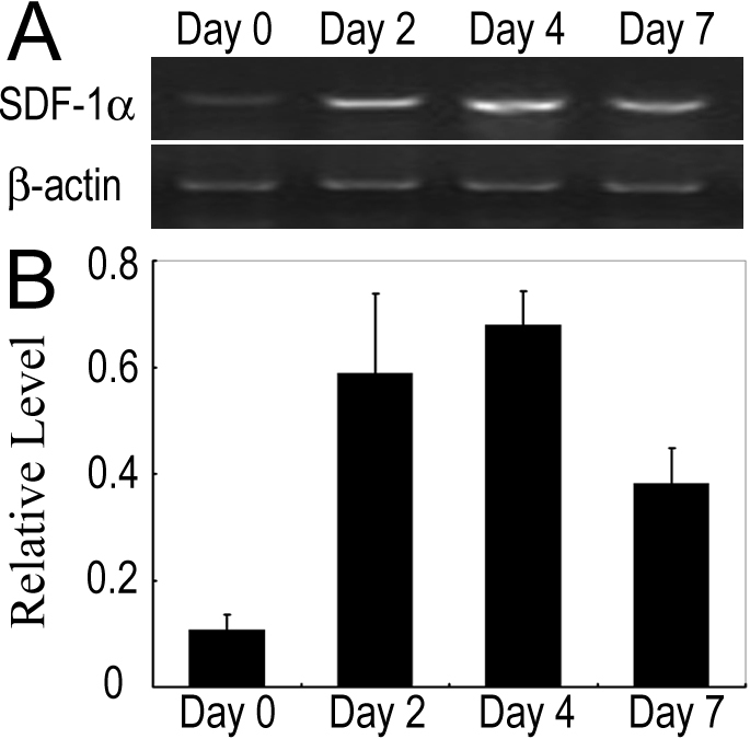
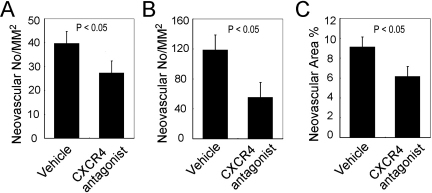
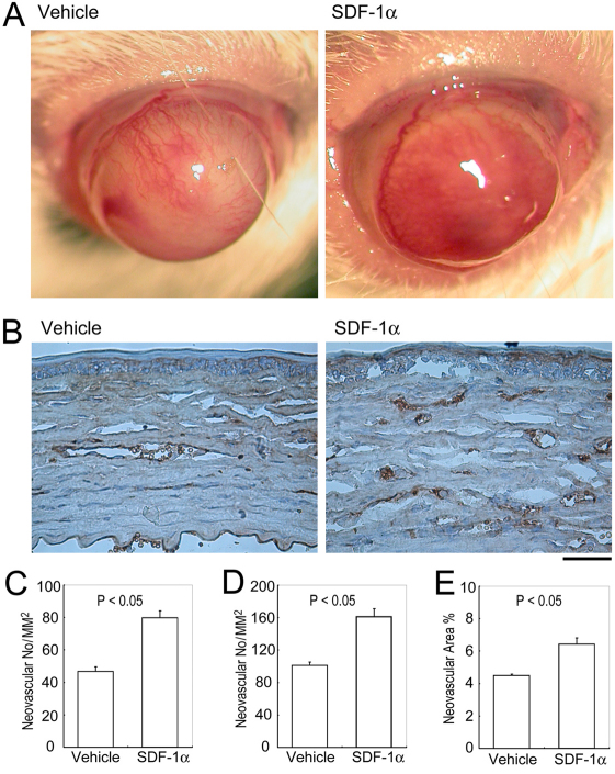
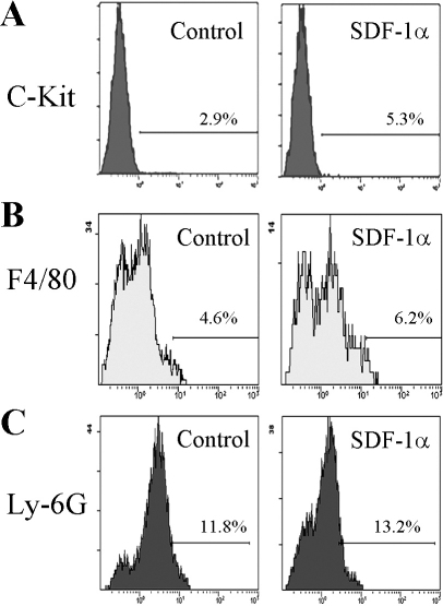
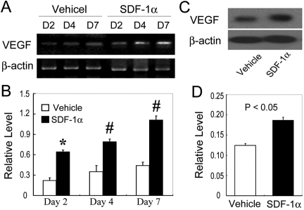
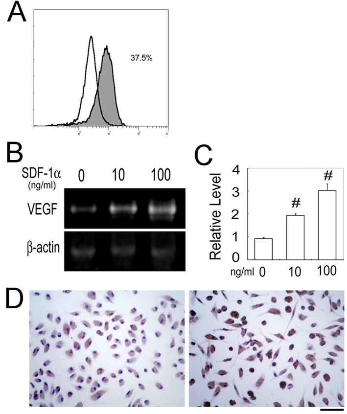
The mRNA expression of SDF-1α was augmented, together with infiltration of c-kit-positive progenitor cells in the corneas after the alkali injury. Compared with vehicle-treated mice, SDF-1α-treated mice exhibited enhanced CNV two weeks after injury, as evidenced by enlarged cluster of differentiation 31 (CD31)-positive areas. Concomitantly, the intracorneal infiltration of c-kit-positive progenitor cells but not F4/80+ macrophages or Ly-6G+ monocytes was significantly enhanced in SDF-1α-treated mice compared to vehicle-treated mice. SDF-1α enhanced vascular endothelial growth factor (VEGF) expression by murine peritoneal macrophages. Enhancement in intraocular VEGF expression was greater in SDF-1α-treated mice than in control mice after injury. Moreover, local administration of C-X-C chemokine receptor type 4 (CXCR4) antagonist after alkali injury reduced alkali-induced CNV.
SDF-1α-treated mice exhibited enhanced alkali-induced CNV through enhanced intracorneal progenitor cell infiltration and increased VEGF expression by macrophages.
探讨基质衍生因子-1(SDF-1)α在实验性角膜新生血管化(CNV)过程中的作用。
通过碱损伤诱导CNV,并在损伤后两周对SDF-1α处理或未处理的小鼠进行比较。损伤后早期血管生成因子的表达通过逆转录聚合酶链反应(RT-PCR)进行定量。损伤后早期祖细胞、巨噬细胞和单核细胞在角膜内的积聚通过流式细胞术分析进行评估。
碱损伤后角膜中SDF-1α的mRNA表达增加,同时c-kit阳性祖细胞浸润。与未处理的小鼠相比,SDF-1α处理的小鼠在损伤后两周表现出增强的CNV,这通过分化簇31(CD31)阳性区域扩大得到证实。同时,与未处理的小鼠相比,SDF-1α处理的小鼠角膜内c-kit阳性祖细胞的浸润显著增强,但F4/80+巨噬细胞或Ly-6G+单核细胞没有增强。SDF-1α增强了小鼠腹腔巨噬细胞血管内皮生长因子(VEGF)的表达。损伤后SDF-1α处理的小鼠眼内VEGF表达的增强比对照小鼠更大。此外,碱损伤后局部给予C-X-C趋化因子受体4(CXCR4)拮抗剂可减少碱诱导的CNV。
SDF-1α处理的小鼠通过增强角膜内祖细胞浸润和巨噬细胞VEGF表达增加,表现出增强的碱诱导的CNV。