Departamento de Química Orgánica Biológica, Instituto de Química Orgánica General, CSIC, 28006 Madrid, Spain.
Nucleic Acids Res. 2011 Dec;39(22):9779-88. doi: 10.1093/nar/gkr667. Epub 2011 Sep 2.
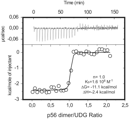
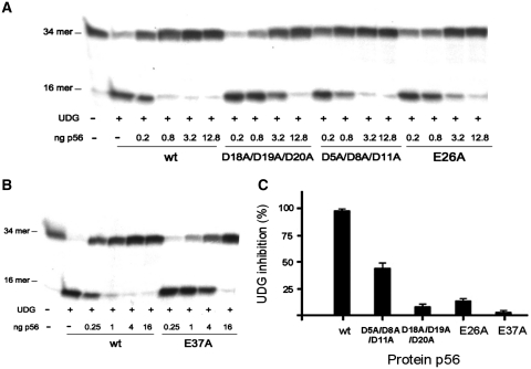
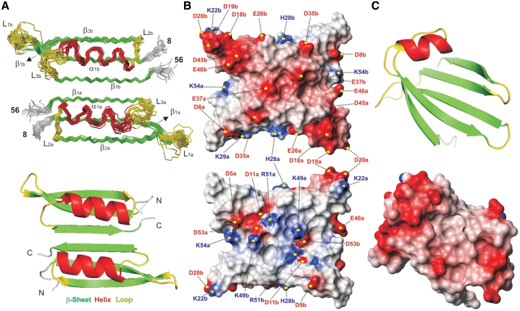
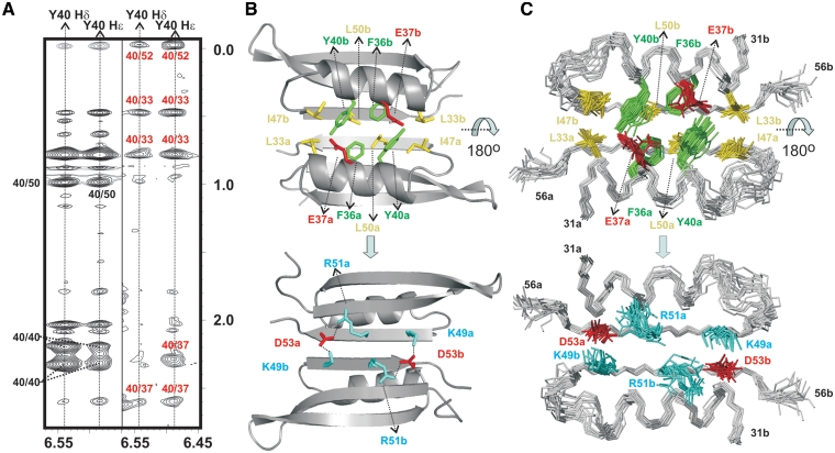
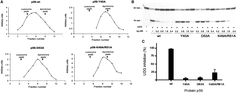
Protein p56 encoded by the Bacillus subtilis phage φ29 inhibits the host uracil-DNA glycosylase (UDG) activity. To get insights into the structural basis for this inhibition, the NMR solution structure of p56 has been determined. The inhibitor defines a novel dimeric fold, stabilized by a combination of polar and extensive hydrophobic interactions. Each polypeptide chain contains three stretches of anti-parallel β-sheets and a helical region linked by three short loops. In addition, microcalorimetry titration experiments showed that it forms a tight 2:1 complex with UDG, strongly suggesting that the dimer represents the functional form of the inhibitor. This was further confirmed by the functional analysis of p56 mutants unable to assemble into dimers. We have also shown that the highly anionic region of the inhibitor plays a significant role in the inhibition of UDG. Thus, based on these findings and taking into account previous results that revealed similarities between the association mode of p56 and the phage PBS-1/PBS-2-encoded inhibitor Ugi with UDG, we propose that protein p56 might inhibit the enzyme by mimicking its DNA substrate.
由枯草芽孢杆菌噬菌体 φ29 编码的蛋白 p56 抑制宿主尿嘧啶-DNA 糖基化酶(UDG)活性。为了深入了解这种抑制的结构基础,已经确定了 p56 的 NMR 溶液结构。该抑制剂定义了一种新的二聚体折叠,由极性和广泛的疏水相互作用组合稳定。每个多肽链包含三个反平行 β-折叠区和一个螺旋区,由三个短环连接。此外,微量热滴定实验表明,它与 UDG 形成紧密的 2:1 复合物,强烈表明二聚体代表抑制剂的功能形式。这通过无法组装成二聚体的 p56 突变体的功能分析得到进一步证实。我们还表明,抑制剂的高度阴离子区域在抑制 UDG 中起着重要作用。因此,基于这些发现,并考虑到先前的结果表明 p56 的结合模式与噬菌体 PBS-1/PBS-2 编码的抑制剂 Ugi 与 UDG 之间存在相似性,我们提出蛋白 p56 可能通过模拟其 DNA 底物来抑制该酶。