Fieselmann Andreas, Kowarschik Markus, Ganguly Arundhuti, Hornegger Joachim, Fahrig Rebecca
Pattern Recognition Lab, Department of Computer Science, Friedrich-Alexander University of Erlangen-Nuremberg, Martensstraße 3, 91058 Erlangen, Germany.
Int J Biomed Imaging. 2011;2011:467563. doi: 10.1155/2011/467563. Epub 2011 Aug 28.
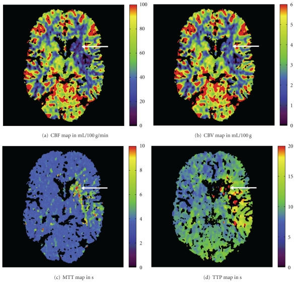
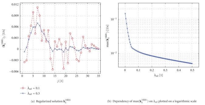
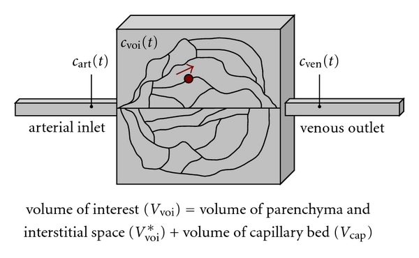
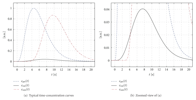
Deconvolution-based analysis of CT and MR brain perfusion data is widely used in clinical practice and it is still a topic of ongoing research activities. In this paper, we present a comprehensive derivation and explanation of the underlying physiological model for intravascular tracer systems. We also discuss practical details that are needed to properly implement algorithms for perfusion analysis. Our description of the practical computer implementation is focused on the most frequently employed algebraic deconvolution methods based on the singular value decomposition. In particular, we further discuss the need for regularization in order to obtain physiologically reasonable results. We include an overview of relevant preprocessing steps and provide numerous references to the literature. We cover both CT and MR brain perfusion imaging in this paper because they share many common aspects. The combination of both the theoretical as well as the practical aspects of perfusion analysis explicitly emphasizes the simplifications to the underlying physiological model that are necessary in order to apply it to measured data acquired with current CT and MR scanners.
基于去卷积的CT和MR脑灌注数据分析在临床实践中被广泛应用,并且它仍然是当前研究活动的一个主题。在本文中,我们对血管内示踪剂系统的基础生理模型进行了全面的推导和解释。我们还讨论了正确实现灌注分析算法所需的实际细节。我们对实际计算机实现的描述集中在基于奇异值分解的最常用代数去卷积方法上。特别是,我们进一步讨论了为获得生理上合理的结果而进行正则化的必要性。我们包括了相关预处理步骤的概述,并提供了大量文献参考。本文涵盖了CT和MR脑灌注成像,因为它们有许多共同之处。灌注分析的理论和实践方面的结合明确强调了为将基础生理模型应用于当前CT和MR扫描仪获取的测量数据而进行的必要简化。