• 文献检索
  • 文档翻译
  • 深度研究
  • 学术资讯
  • Suppr Zotero 插件Zotero 插件
  • 邀请有礼
  • 套餐&价格
  • 历史记录
应用&插件
Suppr Zotero 插件Zotero 插件浏览器插件Mac 客户端Windows 客户端微信小程序
定价
高级版会员购买积分包购买API积分包
服务
文献检索文档翻译深度研究API 文档MCP 服务
关于我们
关于 Suppr公司介绍联系我们用户协议隐私条款
关注我们

Suppr 超能文献

核心技术专利:CN118964589B侵权必究
粤ICP备2023148730 号-1Suppr @ 2026

文献检索

告别复杂PubMed语法,用中文像聊天一样搜索,搜遍4000万医学文献。AI智能推荐,让科研检索更轻松。

立即免费搜索

文件翻译

保留排版,准确专业,支持PDF/Word/PPT等文件格式,支持 12+语言互译。

免费翻译文档

深度研究

AI帮你快速写综述,25分钟生成高质量综述,智能提取关键信息,辅助科研写作。

立即免费体验

术中超声在引导显微神经外科切除小皮质下病变中的应用。

Application of intraoperative ultrasonography for guiding microneurosurgical resection of small subcortical lesions.

机构信息

Department of Ultrasound, Tangdu Hospital of the Fourth Military Medicine University, Xi an 710038, China.

出版信息

Korean J Radiol. 2011 Sep-Oct;12(5):541-6. doi: 10.3348/kjr.2011.12.5.541. Epub 2011 Aug 24.

DOI:10.3348/kjr.2011.12.5.541
PMID:21927554
原文链接:https://pmc.ncbi.nlm.nih.gov/articles/PMC3168794/
Abstract

OBJECTIVE

We wanted to evaluate the clinical value of intraoperative ultrasonography for real-time guidance when performing microneurosurgical resection of small subcortical lesions.

MATERIALS AND METHODS

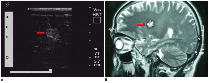
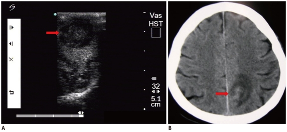
Fifty-two patients with small subcortical lesions were involved in this study. The pathological diagnoses were cavernous hemangioma in 25 cases, cerebral glioma in eight cases, abscess in eight cases, small inflammatory lesion in five cases, brain parasite infection in four cases and the presence of an intracranial foreign body in two cases. An ultrasonic probe was sterilized and lightly placed on the surface of the brain during the operation. The location, extent, characteristics and adjacent tissue of the lesion were observed by high frequency ultrasonography during the operation.

RESULTS

All the lesions were located in the cortex and their mean size was 1.3 ± 0.2 cm. Intraoperative ultrasonography accurately located all the small subcortical lesions, and so the neurosurgeon could provide appropriate treatment. Different lesion pathologies presented with different ultrasonic appearances. Cavernous hemangioma exhibited irregular shapes with distinct margins and it was mildly hyperechoic or hyperechoic. The majority of the cerebral gliomas displayed irregular shapes with indistinct margins, and they often showed cystic and solid mixed echoes. Postoperative imaging identified that the lesions had completely disappeared, and the original symptoms of all the patients were significantly alleviated.

CONCLUSION

Intraoperative ultrasonography can help accurately locate small subcortical lesions and it is helpful for selecting the proper approach and guiding thorough resection of these lesions.

摘要

目的

我们旨在评估术中超声实时引导在施行显微神经外科切除小皮质下病变中的临床价值。

材料与方法

本研究纳入 52 例小皮质下病变患者。病理诊断为海绵状血管瘤 25 例,脑胶质瘤 8 例,脓肿 8 例,小炎性病变 5 例,脑寄生虫感染 4 例,颅内异物 2 例。手术过程中,对超声探头进行消毒后,轻轻放置在脑表面。术中高频超声观察病变的位置、范围、特征及毗邻组织。

结果

所有病变均位于皮质,平均大小为 1.3 ± 0.2cm。术中超声准确定位所有小皮质下病变,使神经外科医生能够提供适当的治疗。不同的病变病理表现出不同的超声表现。海绵状血管瘤呈不规则形状,边界清楚,呈轻度高回声或高回声。大多数脑胶质瘤呈不规则形状,边界不清,常呈囊实性混合回声。术后影像学检查显示病变完全消失,所有患者的原有症状均明显缓解。

结论

术中超声有助于准确定位小皮质下病变,有助于选择合适的手术入路,并指导病变的彻底切除。

https://cdn.ncbi.nlm.nih.gov/pmc/blobs/139a/3168794/6bf63b957276/kjr-12-541-g003.jpg
https://cdn.ncbi.nlm.nih.gov/pmc/blobs/139a/3168794/f56421f9cca7/kjr-12-541-g001.jpg
https://cdn.ncbi.nlm.nih.gov/pmc/blobs/139a/3168794/6bf63b957276/kjr-12-541-g003.jpg
https://cdn.ncbi.nlm.nih.gov/pmc/blobs/139a/3168794/f56421f9cca7/kjr-12-541-g001.jpg
https://cdn.ncbi.nlm.nih.gov/pmc/blobs/139a/3168794/6bf63b957276/kjr-12-541-g003.jpg

相似文献

1
Application of intraoperative ultrasonography for guiding microneurosurgical resection of small subcortical lesions.术中超声在引导显微神经外科切除小皮质下病变中的应用。
Korean J Radiol. 2011 Sep-Oct;12(5):541-6. doi: 10.3348/kjr.2011.12.5.541. Epub 2011 Aug 24.
2
The relationship between intra-operative ultrasonography and pathological grade in cerebral glioma.脑胶质瘤术中超声检查与病理分级的关系
J Int Med Res. 2008 Nov-Dec;36(6):1426-34. doi: 10.1177/147323000803600632.
3
Relationship of intraoperative ultrasound characteristics with pathological grades and Ki-67 proliferation index in intracranial gliomas.颅内胶质瘤术中超声特征与病理分级及Ki-67增殖指数的关系
J Med Ultrason (2001). 2015 Apr;42(2):231-7. doi: 10.1007/s10396-014-0593-1. Epub 2014 Nov 22.
4
[Application of intraoperative ultrasound combined with neuronavigation for resection of intracranial cavernous malformations in minimally invasive neurosurgery].术中超声联合神经导航在微创神经外科手术中切除颅内海绵状畸形的应用
Zhonghua Wai Ke Za Zhi. 2011 Aug 1;49(8):716-9.
5
Intraoperative magnetic resonance imaging for resection of intra-axial brain lesions: a decade of experience using low-field magnetic resonance imaging, Polestar N-10, 20, 30 systems.术中磁共振成像在脑内病变切除中的应用:使用低场磁共振成像、Polestar N-10、30 系统 10 年的经验。
World Neurosurg. 2014 Nov;82(5):770-6. doi: 10.1016/j.wneu.2014.02.004. Epub 2014 Feb 8.
6
[Value of intraoperative ultrasound in precise glioma resection].[术中超声在精准胶质瘤切除中的价值]
Zhonghua Yi Xue Za Zhi. 2013 Sep 3;93(33):2671-3.
7
A simple, safe and effective surface marking and targeting method combined with intraoperative ultrasonography for small subcortical intracranial lesions.一种结合术中超声检查用于小的皮质下颅内病变的简单、安全且有效的表面标记和靶向方法。
J Neurosurg Sci. 2019 Jun;63(3):270-279. doi: 10.23736/S0390-5616.16.03336-1. Epub 2015 Jul 14.
8
Efficacy of intraoperative ultrasonography in neurosurgical tumor resection.术中超声检查在神经外科肿瘤切除术中的疗效
J Neurosurg Pediatr. 2018 May;21(5):504-510. doi: 10.3171/2017.11.PEDS17473. Epub 2018 Feb 16.
9
Intraoperative mapping and monitoring of the corticospinal tracts with neurophysiological assessment and 3-dimensional ultrasonography-based navigation. Clinical article.术中皮质脊髓束的神经生理评估与基于三维超声导航的定位与监测。临床文章。
J Neurosurg. 2011 Mar;114(3):738-46. doi: 10.3171/2010.8.JNS10639. Epub 2010 Aug 27.
10
A practical grading system of ultrasonographic visibility for intracerebral lesions.一种实用的脑内病变超声可见度分级系统。
Acta Neurochir (Wien). 2013 Dec;155(12):2293-8. doi: 10.1007/s00701-013-1868-9. Epub 2013 Sep 13.

引用本文的文献

1
Transcortical transcatheter ultrasound-assisted technique for deep-seated brain tumors. Technical note.经皮质经导管超声辅助技术治疗深部脑肿瘤。技术说明。
J Ultrasound. 2024 Mar;27(1):191-197. doi: 10.1007/s40477-023-00845-w. Epub 2024 Feb 2.
2
Non-enhancing gliomas: does intraoperative ultrasonography improve resections?非强化型胶质瘤:术中超声检查能否提高切除率?
Ultrasonography. 2019 Apr;38(2):156-165. doi: 10.14366/usg.18032. Epub 2018 Jul 29.
3
Intraoperative imaging techniques for glioma surgery.术中成像技术在脑胶质瘤手术中的应用。

本文引用的文献

1
The relationship between intra-operative ultrasonography and pathological grade in cerebral glioma.脑胶质瘤术中超声检查与病理分级的关系
J Int Med Res. 2008 Nov-Dec;36(6):1426-34. doi: 10.1177/147323000803600632.
2
Application of intraoperative ultrasound in neurological surgery.术中超声在神经外科手术中的应用。
Minim Invasive Neurosurg. 2007 Jun;50(3):155-9. doi: 10.1055/s-2007-985146.
3
Ultrasound guidance in intracranial tumor resection: correlation with postoperative magnetic resonance findings.颅内肿瘤切除术中的超声引导:与术后磁共振成像结果的相关性
Future Oncol. 2017 Aug;13(19):1731-1745. doi: 10.2217/fon-2017-0092. Epub 2017 Aug 10.
4
Relationship of intraoperative ultrasound characteristics with pathological grades and Ki-67 proliferation index in intracranial gliomas.颅内胶质瘤术中超声特征与病理分级及Ki-67增殖指数的关系
J Med Ultrason (2001). 2015 Apr;42(2):231-7. doi: 10.1007/s10396-014-0593-1. Epub 2014 Nov 22.
Acta Radiol. 2005 Nov;46(7):743-9. doi: 10.1080/02841850500223208.
4
The guidance of intraoperative ultrasonography in the surgical treatment of arteriovenous malformation.术中超声检查在动静脉畸形外科治疗中的指导作用
Minim Invasive Neurosurg. 2003 Jun;46(3):169-72. doi: 10.1055/s-2003-40737.
5
[Navigation systems for neurosurgery at present and in the future].[当前及未来的神经外科手术导航系统]
No Shinkei Geka. 2003 Jun;31(6):609-18.
6
Intraoperative ultrasound imaging: comparison of pathomorphological findings in US versus CT, MRI and intraoperative findings.术中超声成像:超声与CT、MRI的病理形态学结果及术中结果比较
Acta Neurochir Suppl. 2003;85:95-9. doi: 10.1007/978-3-7091-6043-5_13.
7
Clinical application of proton magnetic resonance spectroscopy in the diagnosis of intracranial mass lesions.质子磁共振波谱在颅内占位性病变诊断中的临床应用
Neuroradiology. 2002 May;44(5):371-81. doi: 10.1007/s00234-001-0760-0. Epub 2002 Feb 21.
8
The echo-guided removal of cerebral cavernous angiomas.超声引导下切除脑海绵状血管瘤。
Acta Neurochir (Wien). 1993;123(3-4):113-7. doi: 10.1007/BF01401865.
9
Transcranial color-coded real-time sonography in the evaluation of intracranial neoplasms and arteriovenous malformations.经颅彩色编码实时超声在颅内肿瘤和动静脉畸形评估中的应用
Neurosurgery. 1992 Sep;31(3):420-8. doi: 10.1227/00006123-199209000-00006.