Soltermann Alex, Kilgus-Hawelski Sandra, Behnke Silvia, Storz Martina, Moch Holger, Bode Beata
Institute of Surgical Pathology, University Hospital Zurich, Schmelzbergstrasse 12, CH-8091 Zurich, Switzerland.
J Clin Bioinforma. 2011 Sep 30;1:25. doi: 10.1186/2043-9113-1-25.
The excision repair cross-complementation group 1 (ERCC1) protein is the key enzyme of the nucleotide excision repair (NER) pathway. Loss of protein expression on immunohistochemistry is predictive for platinum-based chemotherapy response. Frequently, the diagnosis of malignancy is made on cytologic effusion samples. Therefore, we evaluated the staining quality of monoclonal anti-ERCC1 antibodies 8F1 and D-10 on microarrays of malignant pleural and peritoneal effusions by automated immunochemistry.
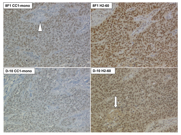
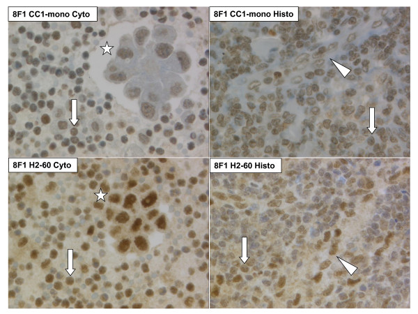
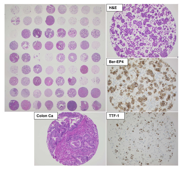
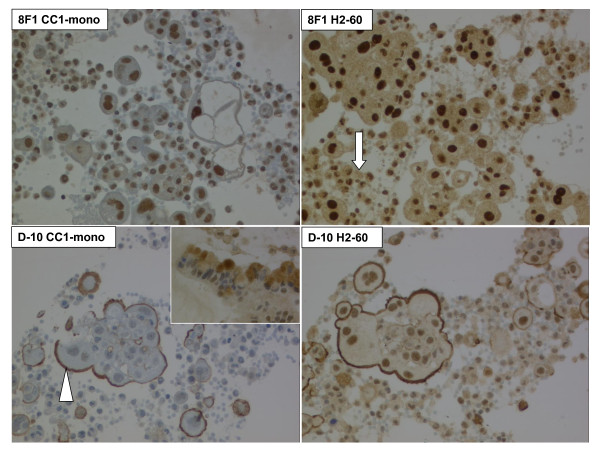
Cores from effusion cell blocks of 117 patients with > 40 malignant cell clusters per whole section (pleural n = 75, peritoneal n = 42) were assembled together with 30 histologic control cores from large tissue blocks (lung, breast and ovarian carcinoma, each n = 10) on hybrid cytology-tissue microarrays (C/TMA). Four immunochemistry protocols (Mab 8F1 and D-10, CC1-mono Ventana and H2-60 Bond automat) were performed. Immunoreactivity was semi-quantitatively scored for intensity and intensity multiplied by percentage staining (H-score).
Tumors were classified into female genital tract carcinoma (n = 39), lung adenocarcinoma (n = 23), mesothelioma (n = 15), unknown primary (n = 14), breast carcinoma (n = 10), gastro-intestinal carcinoma (n = 12) and other (n = 4). On both platforms, reproducible nuclear ERCC1 immunoreactivity was achieved with both antibodies, although D-10 was slightly weaker and presented more background staining as well as more variation in the low expression range. No significant differences were found between cytologic and histologic cores. Using the 8F1 CC1-mono protocol, lung and breast carcinomas had lower ERCC1 expression in comparison to the other entities (p-value < 0.05).
Cytology microarrays (CMA) are suitable for investigation of clinical biomarkers and can be combined with conventional TMA's. Dichotomization of ERCC1 immunoreactivity scores is most suitable for patient stratification since definition of negativity is antibody-dependent.
切除修复交叉互补基因1(ERCC1)蛋白是核苷酸切除修复(NER)途径的关键酶。免疫组织化学检测中蛋白表达缺失可预测铂类化疗反应。恶性肿瘤的诊断常常基于细胞学积液样本。因此,我们通过自动免疫化学方法评估了单克隆抗ERCC1抗体8F1和D-10在恶性胸腔和腹腔积液微阵列上的染色质量。
将117例每整张切片有>40个恶性细胞簇的患者(胸腔积液75例,腹腔积液42例)的积液细胞块的芯与来自大组织块(肺癌、乳腺癌和卵巢癌,各10例)的30个组织学对照芯一起组装到杂交细胞学-组织微阵列(C/TMA)上。执行了四种免疫化学方案(单克隆抗体8F1和D-10、CC1-单克隆Ventana和H2-60 Bond自动染色仪)。免疫反应性根据强度以及强度乘以染色百分比(H评分)进行半定量评分。
肿瘤分为女性生殖道癌(39例)、肺腺癌(23例)、间皮瘤(15例)、原发灶不明(14例)、乳腺癌(10例)、胃肠道癌(12例)和其他(4例)。在两个平台上,两种抗体均实现了可重复的核ERCC1免疫反应性,尽管D-10稍弱,背景染色更多,且在低表达范围内变化更大。细胞学芯和组织学芯之间未发现显著差异。使用8F1 CC1-单克隆方案时,与其他实体相比,肺癌和乳腺癌的ERCC1表达较低(p值<0.05)。
细胞学微阵列(CMA)适用于临床生物标志物的研究,并且可以与传统的组织微阵列相结合。ERCC1免疫反应性评分的二分法最适合患者分层,因为阴性的定义依赖于抗体。