Jantaratnotai Nattinee, Ryu Jae K, Schwab Claudia, McGeer Patrick L, McLarnon James G
Department of Anesthesiology, Pharmacology and Therapeutics, Faculty of Medicine, The University of British Columbia, 2176 Health Sciences Mall, Vancouver, BC, V6T 1Z3, Canada.
Int J Alzheimers Dis. 2011;2011:918280. doi: 10.4061/2011/918280. Epub 2011 Sep 29.
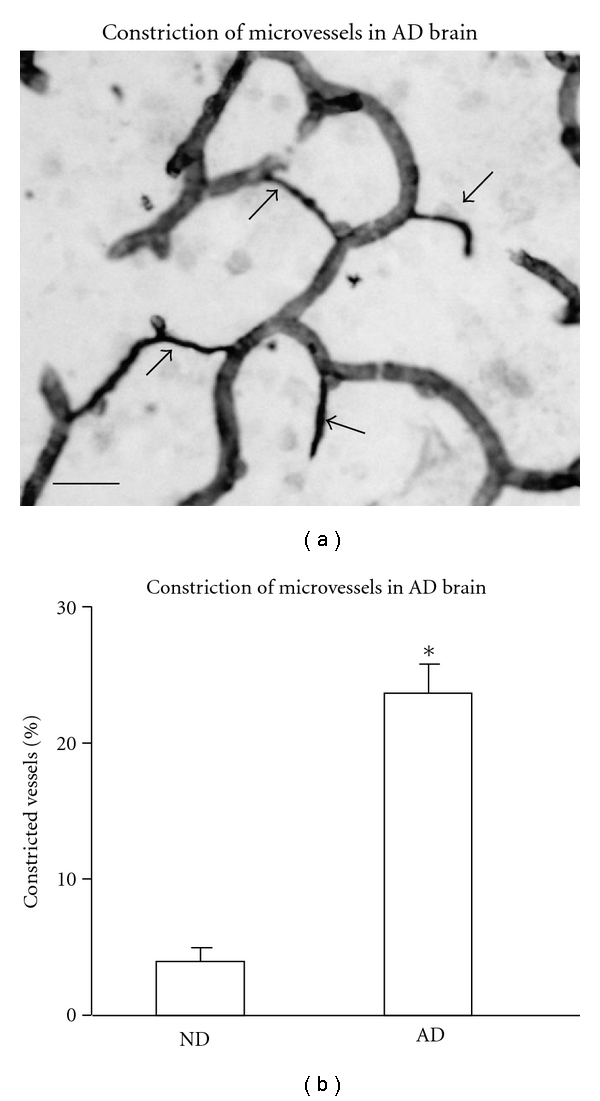
The validity of amyloid-β peptide (Aβ(1-42)) intrahippocampal injection, as an animal model of Alzheimer's disease (AD), has previously been considered in terms of inflammatory reactivity and neuronal damage. In this work, we have extended the testing of the animal model to vasculature by comparison of selected properties of microvessels in vivo with those in human AD brain tissue. The injection of Aβ(1-42), relative to control PBS (phosphate buffered saline), increased the mean number of microvessels and diminished the mean length of microvessels in the molecular layer of dentate gyrus. The animal model showed Aβ(1-42), but not PBS, injection was associated with abnormalities in morphology of microvessels which were characterized as looping, fragmented, knob-like, uneven, and constricted. In particular, numbers of constricted microvessels, defined as vessels with diameters less than 3 μm, were considerably enhanced for Aβ(1-42), compared to PBS, injection. In comparison, human AD brain demonstrated an elevated number of microvessels with a diminished mean length relative to nondemented (ND) brain. Additionally, microvessel perturbations in AD brain showed a similar pattern of morphological abnormalities to those observed in Aβ(1-42)-injected rat hippocampus. Constricted microvessels were a prominent feature of AD brain but were rarely observed in ND tissue. These results provide the first evidence that a peptide-injection animal model exhibits a commonality in perturbations of microvessels compared with those evident in AD brain.
淀粉样β肽(Aβ(1 - 42))海马体内注射作为阿尔茨海默病(AD)的动物模型,其有效性先前已根据炎症反应性和神经元损伤进行过考量。在本研究中,我们通过比较体内微血管的选定特性与人类AD脑组织中的特性,将该动物模型的测试扩展至脉管系统。相对于对照磷酸盐缓冲盐水(PBS),注射Aβ(1 - 42)增加了齿状回分子层中微血管的平均数量,并缩短了微血管的平均长度。该动物模型显示,注射Aβ(1 - 42)而非PBS与微血管形态异常有关,这些异常表现为成环、断裂、瘤状、不均匀和狭窄。特别是,定义为直径小于3μm的狭窄微血管数量,与注射PBS相比,注射Aβ(1 - 42)后显著增加。相比之下,人类AD脑显示微血管数量增加,相对于非痴呆(ND)脑平均长度缩短。此外,AD脑中微血管的扰动显示出与在注射Aβ(1 - 42)的大鼠海马体中观察到的形态异常相似的模式。狭窄微血管是AD脑的一个突出特征,但在ND组织中很少观察到。这些结果提供了首个证据,表明肽注射动物模型在微血管扰动方面与AD脑中明显的情况具有共性。