Cukras Catherine A, Wong Wai T, Caruso Rafael, Cunningham Denise, Zein Wadih, Sieving Paul A
Division of Epidemiology and Clinical Research, National Eye Institute, National Institutes of Health, Bethesda, MD 20892, USA.
Arch Ophthalmol. 2012 Feb;130(2):171-9. doi: 10.1001/archophthalmol.2011.332. Epub 2011 Oct 10.
To study the longitudinal changes in autofluorescence in Stargardt disease to reveal aspects of disease progression not previously evident. Changes in autofluorescence reflect changing fluorophore compositions of lipofuscin and melanin in retinal pigment epithelial cells, which has been hypothesized to contribute to Stargardt disease pathogenesis.
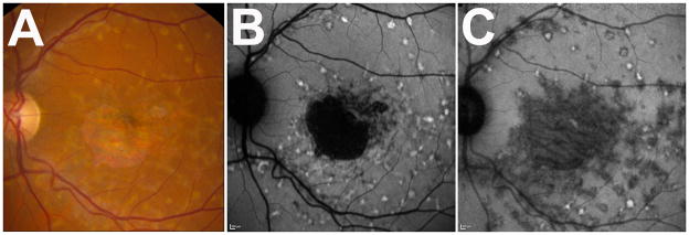
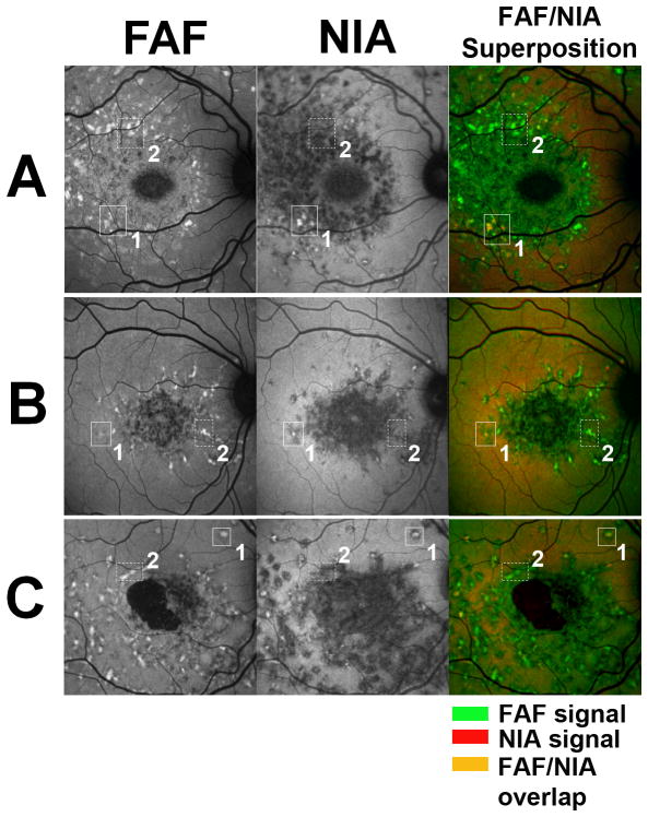
We examined the temporospatial patterns of fundus autofluorescence with excitation at both 488 nm (standard fundus autofluorescence) and 795 nm (near-infrared autofluorescence) in a longitudinal case series involving 8 eyes of 4 patients (range of follow-up, 11-57 months; mean, 39 months). Image processing was performed to analyze spatial and temporal cross-modality associations.
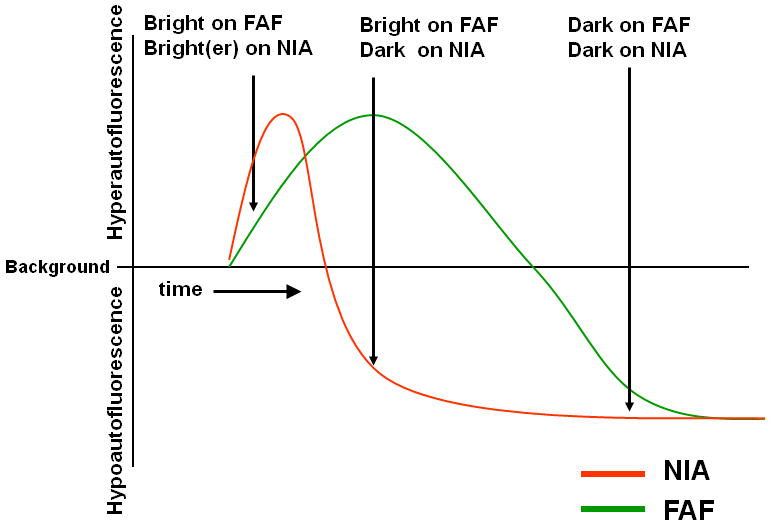
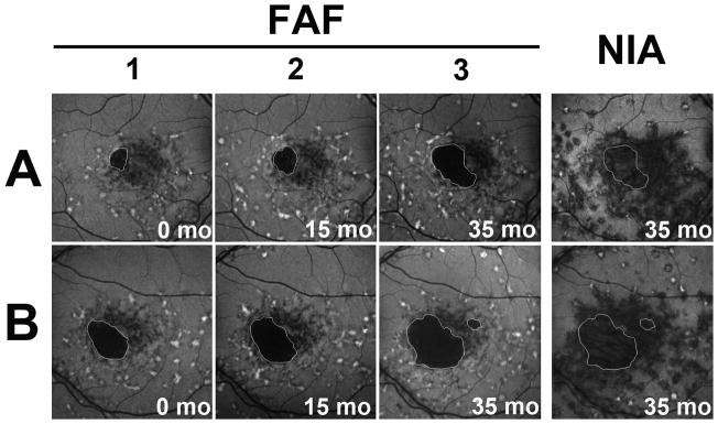
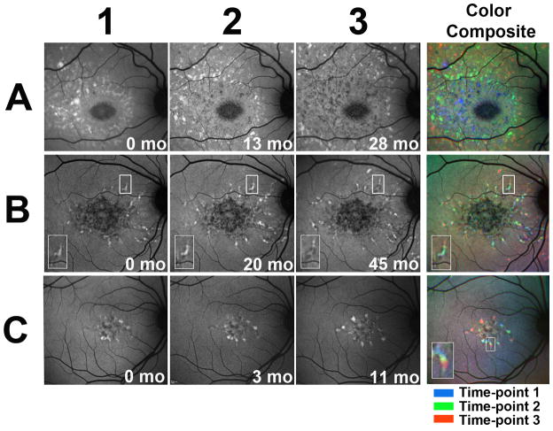
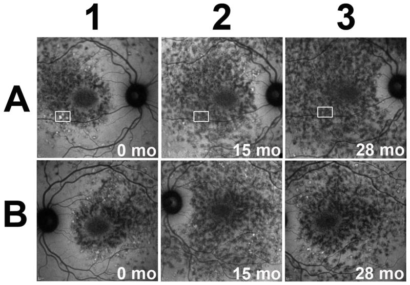
Longitudinal fundus autofluorescence imaging of fleck lesions revealed hyperautofluorescent lesions that extended in a centrifugal direction from the fovea with time. Patterns of spread were nonrandom and followed a radial path that left behind a trail of diminishing autofluorescence. Longitudinal near-infrared autofluorescence imaging also demonstrated centrifugal lesion spread but with fewer hyperautofluorescent lesions, suggestive of more transient hyperautofluorescence and more rapid decay at longer wavelengths. Fundus autofluorescence and near-infrared autofluorescence abnormalities were spatially correlated with each other, and together they reflect systematic progressions in fleck distribution and fluorophore composition occurring during the natural history of the disease.
Stargardt disease fleck lesions do not evolve randomly in location but instead follow consistent patterns of radial expansion and a systematic decay of autofluorescence that reflect changing lipofuscin and melanin compositions in retinal pigment epithelial cells. These progressive foveal-to-peripheral changes are helpful in elucidating molecular and cellular mechanisms underlying Stargardt disease and may constitute potential outcome measures in clinical trials.
研究斯塔加特病中自发荧光的纵向变化,以揭示此前未被发现的疾病进展情况。自发荧光的变化反映了视网膜色素上皮细胞中脂褐素和黑色素荧光团组成的变化,这被认为与斯塔加特病的发病机制有关。
我们对4例患者的8只眼睛进行了纵向病例系列研究,在488nm(标准眼底自发荧光)和795nm(近红外自发荧光)激发下检查眼底自发荧光的时空模式(随访时间范围为11 - 57个月;平均39个月)。进行图像处理以分析空间和时间上的跨模态关联。
对斑点状病变的纵向眼底自发荧光成像显示,随着时间推移,高自发荧光病变从中央凹向周边呈离心方向扩展。扩展模式并非随机,而是沿着一条径向路径,留下自发荧光逐渐减弱的痕迹。纵向近红外自发荧光成像也显示病变呈离心扩展,但高自发荧光病变较少,提示在较长波长下高自发荧光更短暂且衰减更快。眼底自发荧光和近红外自发荧光异常在空间上相互关联,共同反映了疾病自然史中斑点分布和荧光团组成的系统性进展。
斯塔加特病的斑点状病变并非在位置上随机演变,而是遵循一致的径向扩展模式和自发荧光的系统性衰减,这反映了视网膜色素上皮细胞中脂褐素和黑色素组成的变化。这些从中央凹到周边的渐进性变化有助于阐明斯塔加特病的分子和细胞机制,可能构成临床试验中的潜在疗效指标。