• 文献检索
  • 文档翻译
  • 深度研究
  • 学术资讯
  • Suppr Zotero 插件Zotero 插件
  • 邀请有礼
  • 套餐&价格
  • 历史记录
应用&插件
Suppr Zotero 插件Zotero 插件浏览器插件Mac 客户端Windows 客户端微信小程序
定价
高级版会员购买积分包购买API积分包
服务
文献检索文档翻译深度研究API 文档MCP 服务
关于我们
关于 Suppr公司介绍联系我们用户协议隐私条款
关注我们

Suppr 超能文献

核心技术专利:CN118964589B侵权必究
粤ICP备2023148730 号-1Suppr @ 2026

文献检索

告别复杂PubMed语法,用中文像聊天一样搜索,搜遍4000万医学文献。AI智能推荐,让科研检索更轻松。

立即免费搜索

文件翻译

保留排版,准确专业,支持PDF/Word/PPT等文件格式,支持 12+语言互译。

免费翻译文档

深度研究

AI帮你快速写综述,25分钟生成高质量综述,智能提取关键信息,辅助科研写作。

立即免费体验

肾脏纤维化的细胞和分子机制。

Cellular and molecular mechanisms of renal fibrosis.

机构信息

Department of Pathology, University of Pittsburgh, S-405 Biomedical Science Tower, 200 Lothrop Street, Pittsburgh, PA 15261, USA.

出版信息

Nat Rev Nephrol. 2011 Oct 18;7(12):684-96. doi: 10.1038/nrneph.2011.149.

DOI:10.1038/nrneph.2011.149
PMID:22009250
原文链接:https://pmc.ncbi.nlm.nih.gov/articles/PMC4520424/
Abstract

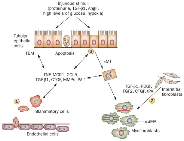
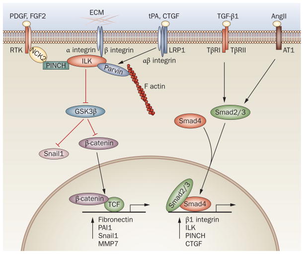
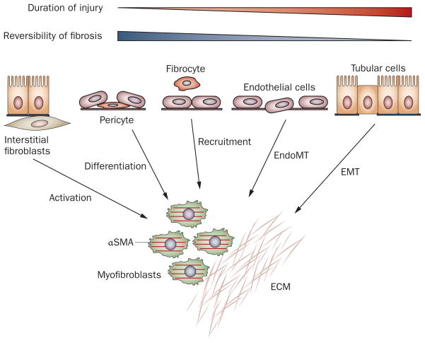
Renal fibrosis, particularly tubulointerstitial fibrosis, is the common final outcome of almost all progressive chronic kidney diseases. Renal fibrosis is also a reliable predictor of prognosis and a major determinant of renal insufficiency. Irrespective of the initial causes, renal fibrogenesis is a dynamic and converging process that consists of four overlapping phases: priming, activation, execution and progression. Nonresolving inflammation after a sustained injury sets up the fibrogenic stage (priming) and triggers the activation and expansion of matrix-producing cells from multiple sources through diverse mechanisms, including activation of interstitial fibroblasts and pericytes, phenotypic conversion of tubular epithelial and endothelial cells and recruitment of circulating fibrocytes. Upon activation, matrix-producing cells assemble a multicomponent, integrin-associated protein complex that integrates input from various fibrogenic signals and orchestrates the production of matrix components and their extracellular assembly. Multiple cellular and molecular events, such as tubular atrophy, microvascular rarefaction and tissue hypoxia, promote scar formation and ensure a vicious progression to end-stage kidney failure. This Review outlines our current understanding of the cellular and molecular mechanisms of renal fibrosis, which could offer novel insights into the development of new therapeutic strategies.

摘要

肾纤维化,特别是肾小管间质纤维化,是几乎所有进行性慢性肾脏病的共同终末结局。肾纤维化也是预后的可靠预测指标,也是肾功能不全的主要决定因素。无论最初的原因是什么,肾纤维化是一个动态和收敛的过程,由四个重叠的阶段组成:启动、激活、执行和进展。持续损伤后的非解决炎症引发纤维化阶段(启动),并通过多种机制触发基质产生细胞的激活和扩张,这些机制包括间质成纤维细胞和周细胞的激活、管状上皮和内皮细胞的表型转化以及循环纤维细胞的募集。激活后,基质产生细胞组装一个多成分、整合素相关蛋白复合物,整合来自各种纤维化信号的输入,并协调基质成分的产生及其细胞外组装。多种细胞和分子事件,如肾小管萎缩、微血管稀疏和组织缺氧,促进瘢痕形成,并确保向终末期肾衰竭的恶性进展。这篇综述概述了我们对肾纤维化的细胞和分子机制的理解,这可能为新的治疗策略的发展提供新的见解。

https://cdn.ncbi.nlm.nih.gov/pmc/blobs/0b8e/4520424/13e2807b53a5/nihms710434f3.jpg
https://cdn.ncbi.nlm.nih.gov/pmc/blobs/0b8e/4520424/fdcc70ea04e5/nihms710434f1.jpg
https://cdn.ncbi.nlm.nih.gov/pmc/blobs/0b8e/4520424/2132f710e827/nihms710434f2.jpg
https://cdn.ncbi.nlm.nih.gov/pmc/blobs/0b8e/4520424/13e2807b53a5/nihms710434f3.jpg
https://cdn.ncbi.nlm.nih.gov/pmc/blobs/0b8e/4520424/fdcc70ea04e5/nihms710434f1.jpg
https://cdn.ncbi.nlm.nih.gov/pmc/blobs/0b8e/4520424/2132f710e827/nihms710434f2.jpg
https://cdn.ncbi.nlm.nih.gov/pmc/blobs/0b8e/4520424/13e2807b53a5/nihms710434f3.jpg

相似文献

1
Cellular and molecular mechanisms of renal fibrosis.肾脏纤维化的细胞和分子机制。
Nat Rev Nephrol. 2011 Oct 18;7(12):684-96. doi: 10.1038/nrneph.2011.149.
2
Renal fibrosis: an update.肾纤维化:最新进展
Curr Opin Nephrol Hypertens. 2001 May;10(3):315-20. doi: 10.1097/00041552-200105000-00004.
3
Cellular and molecular pathways that lead to progression and regression of renal fibrogenesis.导致肾纤维化进展和消退的细胞与分子途径。
Curr Mol Med. 2005 Aug;5(5):467-74. doi: 10.2174/1566524054553478.
4
The origin of renal fibroblasts/myofibroblasts and the signals that trigger fibrosis.肾成纤维细胞/肌成纤维细胞的起源以及引发纤维化的信号。
Differentiation. 2016 Sep;92(3):102-107. doi: 10.1016/j.diff.2016.05.008. Epub 2016 Jun 1.
5
Tenascin is an ubiquitous extracellular matrix protein of human renal interstitium in normal and pathologic conditions.腱生蛋白是一种在正常和病理条件下均存在于人类肾间质中的普遍存在的细胞外基质蛋白。
Nephron. 1996;72(4):579-86. doi: 10.1159/000188943.
6
Renal interstitial fibrosis: a critical evaluation of the origin of myofibroblasts.肾间质纤维化:对肌成纤维细胞起源的批判性评估。
Contrib Nephrol. 2011;169:73-93. doi: 10.1159/000313946. Epub 2011 Jan 20.
7
Contribution of genetics and epigenetics to progression of kidney fibrosis.遗传学和表观遗传学对肾纤维化进展的作用。
Nephrol Dial Transplant. 2014 Sep;29 Suppl 4:iv72-9. doi: 10.1093/ndt/gft025. Epub 2013 Aug 23.
8
Progression of chronic kidney disease: too much cellular talk causes damage.慢性肾脏病的进展:过多的细胞间交流导致损伤。
Kidney Int. 2017 Mar;91(3):552-560. doi: 10.1016/j.kint.2016.08.025. Epub 2016 Oct 20.
9
Janus-Faced: Molecular Mechanisms and Versatile Nature of Renal Fibrosis.两面神:肾脏纤维化的分子机制和多功能性质。
Kidney360. 2020 May 15;1(7):697-704. doi: 10.34067/KID.0001972020. eCollection 2020 Jul 30.
10
Cellular mechanisms of tissue fibrosis. 3. Novel mechanisms of kidney fibrosis.细胞组织纤维化的机制。3. 肾脏纤维化的新机制。
Am J Physiol Cell Physiol. 2013 Apr 1;304(7):C591-603. doi: 10.1152/ajpcell.00414.2012. Epub 2013 Jan 16.

引用本文的文献

1
Therapeutic potential of Desmodium styracifolium polysaccharide in attenuating nano-calcium oxalate induced renal injury and fibrosis.广金钱草多糖在减轻纳米草酸钙诱导的肾损伤和纤维化方面的治疗潜力
Commun Biol. 2025 Sep 2;8(1):1330. doi: 10.1038/s42003-025-08757-7.
2
Quantifying "Medical Renal Disease": A Pediatric Pilot Study Using Ultrasound Radiomics for Differentiating Acute Kidney Injury and Chronic Kidney Disease.量化“医学性肾病”:一项利用超声放射组学鉴别急性肾损伤和慢性肾病的儿科试点研究
Diagnostics (Basel). 2025 Aug 21;15(16):2112. doi: 10.3390/diagnostics15162112.
3
Risk Factors of Histopathological Crescent Formation in Pediatric IgA Vasculitis Nephritis.

本文引用的文献

1
Homeostatic and pathogenic role of renal dendritic cells.肾脏树突状细胞的稳态和致病作用。
Kidney Int. 2011 Jul;80(2):139-45. doi: 10.1038/ki.2011.129. Epub 2011 May 25.
2
The PDGF family in renal fibrosis.血小板衍生生长因子家族在肾纤维化中的作用。
Pediatr Nephrol. 2012 Jul;27(7):1041-50. doi: 10.1007/s00467-011-1892-z. Epub 2011 May 21.
3
An orally active chemokine receptor CCR2 antagonist prevents glomerulosclerosis and renal failure in type 2 diabetes.一种口服活性趋化因子受体 CCR2 拮抗剂可预防 2 型糖尿病肾小球硬化和肾衰竭。
儿童IgA血管炎肾病组织病理学新月体形成的危险因素
Medicina (Kaunas). 2025 Aug 6;61(8):1421. doi: 10.3390/medicina61081421.
4
A High-Fat Diet Increases Kidney Fibrosis Through Regulating TGF-β and PDGF-β Signaling Pathways in Normotensive and Hypertensive Rat Models.高脂饮食通过调节正常血压和高血压大鼠模型中的TGF-β和PDGF-β信号通路增加肾纤维化。
Int J Mol Sci. 2025 Aug 20;26(16):8031. doi: 10.3390/ijms26168031.
5
Exploring the Proteomic Signature of Diabetic Nephropathy: Implications for Early Diagnosis and Treatment.探索糖尿病肾病的蛋白质组学特征:对早期诊断和治疗的意义。
Life (Basel). 2025 Aug 19;15(8):1312. doi: 10.3390/life15081312.
6
A Rare Case Report of Wunderlich Syndrome in a Chronic Hemodialysis Patient.一名慢性血液透析患者的肾自发性破裂综合征罕见病例报告。
Reports (MDPI). 2025 Jul 25;8(3):121. doi: 10.3390/reports8030121.
7
Association of renal cortical thickness and renal function in patients with malignant hypertension with thrombotic microangiopathy: a biopsy-based cohort study from China.恶性高血压合并血栓性微血管病患者肾皮质厚度与肾功能的相关性:一项来自中国的基于活检的队列研究。
BMJ Open. 2025 Aug 16;15(8):e096581. doi: 10.1136/bmjopen-2024-096581.
8
NRF2 Dysregulation and Therapeutic Insights Across Chronic Kidney Diseases.慢性肾脏病中的NRF2失调与治疗见解
Int J Mol Sci. 2025 Aug 2;26(15):7471. doi: 10.3390/ijms26157471.
9
Mechanistic Insights into the Pathogenesis of Polycystic Kidney Disease.多囊肾病发病机制的机制性见解
Cells. 2025 Aug 5;14(15):1203. doi: 10.3390/cells14151203.
10
Fibrosis-4 index as a predictor of all-cause and cardiovascular mortality in patients with chronic kidney disease.纤维化-4指数作为慢性肾脏病患者全因死亡率和心血管死亡率的预测指标
PLoS One. 2025 Aug 1;20(8):e0329315. doi: 10.1371/journal.pone.0329315. eCollection 2025.
Kidney Int. 2011 Jul;80(1):68-78. doi: 10.1038/ki.2011.102. Epub 2011 Apr 20.
4
Chemokines in renal injury.趋化因子在肾损伤中的作用。
J Am Soc Nephrol. 2011 May;22(5):802-9. doi: 10.1681/ASN.2010050510. Epub 2011 Apr 7.
5
Epithelial to mesenchymal transition as a biomarker in renal fibrosis: are we ready for the bedside?上皮-间质转化作为肾纤维化的生物标志物:我们是否已准备好应用于临床?
Fibrogenesis Tissue Repair. 2011 Apr 6;4:11. doi: 10.1186/1755-1536-4-11.
6
SnapShot: The epithelial-mesenchymal transition.简讯:上皮-间质转化
Cell. 2011 Apr 1;145(1):162.e1. doi: 10.1016/j.cell.2011.03.029.
7
Scar wars: mapping the fate of epithelial-mesenchymal-myofibroblast transition.瘢痕战争:绘制上皮-间充质-肌成纤维细胞转化的命运图。
Kidney Int. 2011 Jul;80(1):41-50. doi: 10.1038/ki.2011.77. Epub 2011 Mar 23.
8
Mechanisms of fibrosis: the role of the pericyte.纤维化机制:周细胞的作用。
Curr Opin Nephrol Hypertens. 2011 May;20(3):297-305. doi: 10.1097/MNH.0b013e328344c3d4.
9
Fibrosis and evidence for epithelial-mesenchymal transition in the kidneys of patients with staghorn calculi.肾结石患者肾脏中的纤维化和上皮-间充质转化的证据。
BJU Int. 2011 Oct;108(8):1336-45. doi: 10.1111/j.1464-410X.2010.10074.x. Epub 2011 Mar 16.
10
A microRNA circuit mediates transforming growth factor-β1 autoregulation in renal glomerular mesangial cells.微小 RNA 电路介导转化生长因子-β1 在肾肾小球系膜细胞中的自身调控。
Kidney Int. 2011 Aug;80(4):358-68. doi: 10.1038/ki.2011.43. Epub 2011 Mar 9.