Department of Pharmacology and Toxicology, NTR College of Veterinary Science, Gannavaram - 521 102, Krishna (Dt), Andhra Pradesh, India.
Indian J Pharmacol. 2011 Sep;43(5):568-73. doi: 10.4103/0253-7613.84974.
The aim of the present study was to investigate whether Tribulus terrestris Linn (TT) could protect the cadmium (Cd)-induced testicular tissue peroxidation in rats and to explore the underlying mechanism of the same.
In vitro and in vivo studies were conducted to know the protective effect of ethanolic extract of TT (eTT) in Cd toxicity. In in vitro studies, total antioxidant and ferrous metal ion chelating activity of TT was studied. In vivo studies were conducted in rats. A total of 40 Wistar strain adult male rats were divided into four groups. Group 1 served as control, while group 2 to 4 received CdCl(2) (3 mg/kg b. wt. s/c once a week). In addition to Cd, group 3 and 4 rats also received eTT (5 mg/kg b.wt. daily as oral gavage) and α-tocopherol (75 mg/kg daily by oral gavage), respectively. At the end of 6(th) week, all the rats were sacrificed and the separated testes were weighted and processed for estimation of tissue peroxidation markers, antioxidant markers, functional markers, and Cd concentration. The testes were also subjected to histopathological screening.
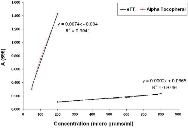
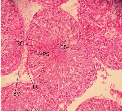
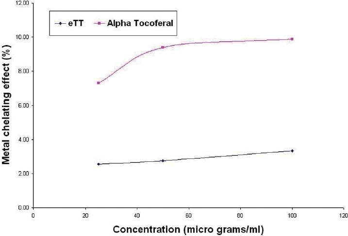
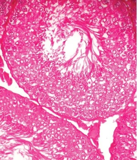
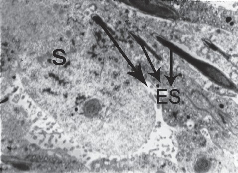
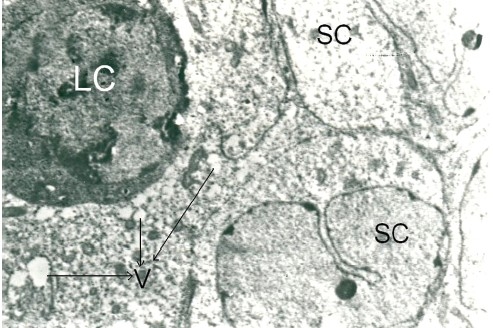
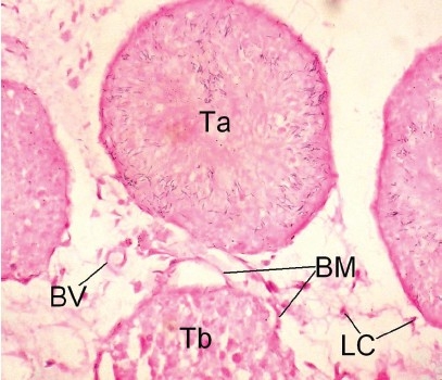
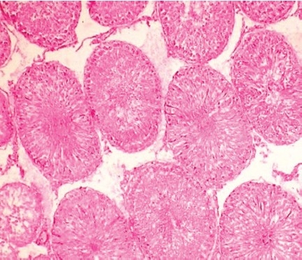
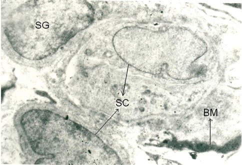
In in vitro studies, the percentage of metal ion chelating activity of 50 μg/ml of eTT and α-tocopherol were 2.76 and 9.39, respectively, and the antioxidant capacity of eTT was equivalent to 0.063 μg of α-tocopherol/μg of eTT. In in vivo studies, administration of Cd significantly reduced the absolute and relative testicular weight, antioxidant markers such as superoxide dismutase and glutathione, and functional markers such as LDH and ALP, along with significant increase in peroxidation markers such as malondialdehyde and protein carbonyls in testicular tissue. Testes of Cd only-treated group showed histological insults like necrotic changes in seminiferous tubules and interstitium, shrunken tubules with desquamated basal lamina, vacuolization and destruction of sertoli cells, and degenerating Leydig cells. This group also had higher Cd levels in testicular tissue. Co-treatment with eTT and α-tocopherol significantly reduced the Cd burden in the testes along with reversal of the Cd-induced changes.
eTT exhibited protective effect against Cd-induced testicular damage. The protective effect appears to be mediated through inhibition of testicular tissue peroxidation by antioxidant and metal chelator activity and also, may be indirectly by stimulating the testosterone production from Leydig cells.
本研究旨在探讨蒺藜(TT)是否能保护镉(Cd)诱导的大鼠睾丸组织过氧化,并探讨其潜在机制。
进行了体外和体内研究,以了解 TT 乙醇提取物(eTT)在 Cd 毒性中的保护作用。在体外研究中,研究了 TT 的总抗氧化和亚铁金属离子螯合活性。体内研究在 Wistar 成年雄性大鼠中进行。将 40 只大鼠分为 4 组。第 1 组为对照组,第 2 至 4 组大鼠每周腹腔注射一次 CdCl2(3mg/kg 体重)。除了 Cd 之外,第 3 和 4 组大鼠还分别给予 eTT(5mg/kg 体重,每日口服灌胃)和 α-生育酚(75mg/kg 体重,每日口服灌胃)。在第 6 周结束时,所有大鼠被处死,分离的睾丸称重,并进行组织过氧化标志物、抗氧化标志物、功能标志物和 Cd 浓度的测定。还对睾丸进行了组织病理学筛查。
在体外研究中,50μg/ml 的 eTT 和 α-生育酚的金属离子螯合活性百分比分别为 2.76%和 9.39%,eTT 的抗氧化能力相当于 0.063μg α-生育酚/μg eTT。在体内研究中,Cd 的给药显著降低了睾丸的绝对和相对重量、超氧化物歧化酶和谷胱甘肽等抗氧化标志物以及 LDH 和 ALP 等功能标志物,同时睾丸组织中的过氧化标志物如丙二醛和蛋白质羰基明显增加。仅给予 Cd 的大鼠睾丸组织出现了坏死性变化,如曲细精管和间质的坏死、基底膜脱落的萎缩小管、空泡化和支持细胞破坏以及退化的 Leydig 细胞。该组睾丸组织中的 Cd 含量也较高。eTT 和 α-生育酚的联合治疗显著降低了睾丸中的 Cd 负荷,并逆转了 Cd 诱导的变化。
eTT 对 Cd 诱导的睾丸损伤具有保护作用。这种保护作用可能是通过抗氧化和金属螯合活性抑制睾丸组织过氧化,以及可能通过刺激 Leydig 细胞产生睾酮而间接实现的。