Department of Obstetrics and Gynecology, School of Medicine, Washington University, 4566 Scott Avenue, St Louis, MO 63110, USA.
Reproduction. 2012 Jan 1;143(1):107-21. doi: 10.1530/REP-11-0340. Epub 2011 Nov 1.
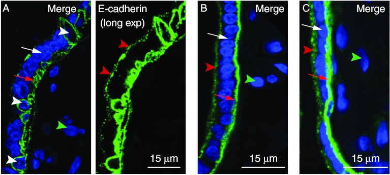
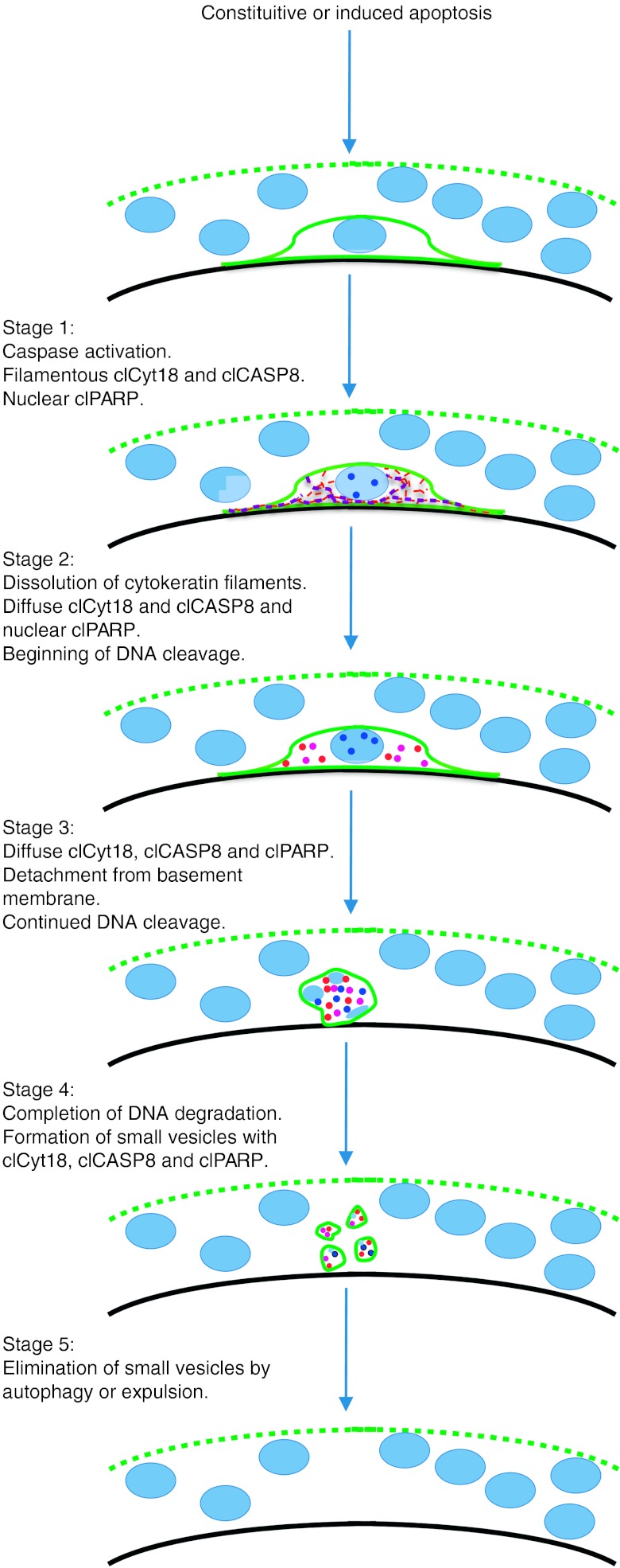
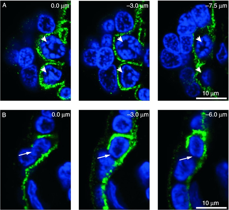
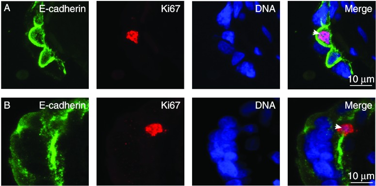
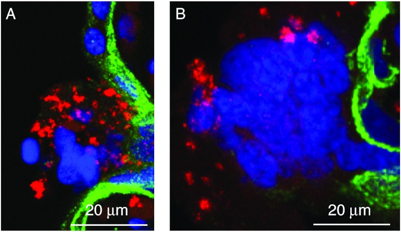
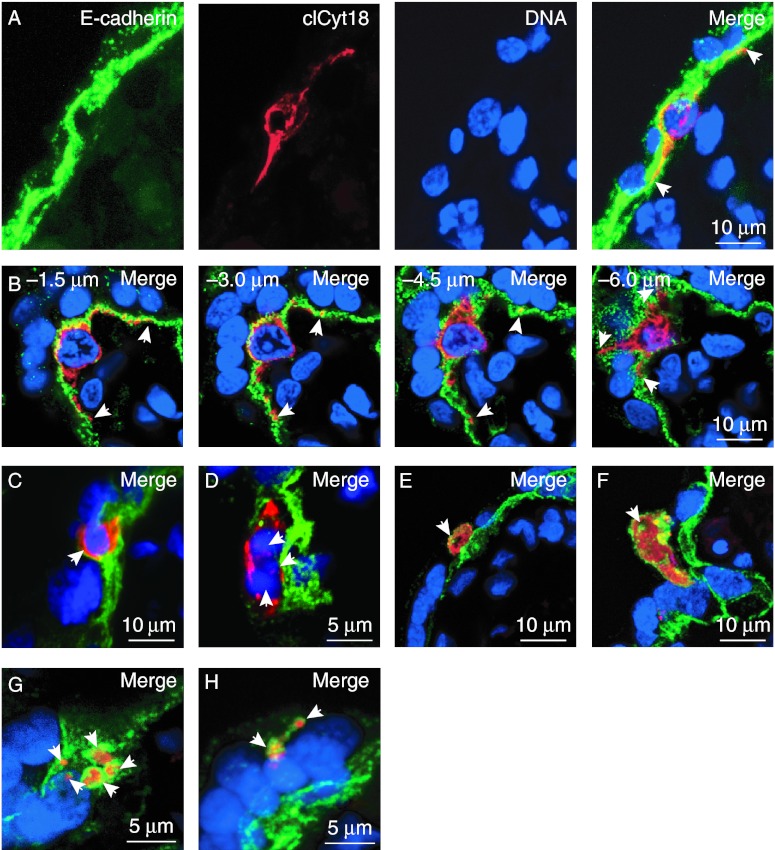
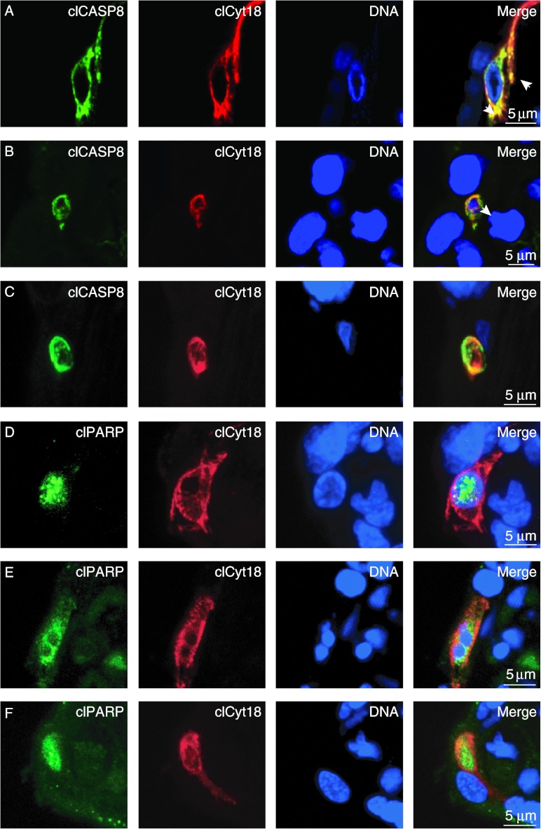
Human placental villi are surfaced by a multinucleated and terminally differentiated epithelium, the syncytiotrophoblast, with a subjacent layer of mononucleated cytotrophoblasts that can divide and fuse to replenish the syncytiotrophoblast. The objectives of this study were i) to develop an approach to definitively identify and distinguish cytotrophoblasts from the syncytiotrophoblast, ii) to unambiguously determine the relative susceptibility of villous cytotrophoblasts and syncytiotrophoblast to constitutive and stress-induced apoptosis mediated by caspases, and iii) to understand the progression of apoptosis in villous trophoblasts. Confocal microscopy with co-staining for E-cadherin and DNA allowed us to clearly distinguish the syncytiotrophoblast from cytotrophoblasts and identified that many cytotrophoblasts are deeply interdigitated into the syncytiotrophoblast. Staining for specific markers of caspase-mediated apoptosis indicate that apoptosis occurs readily in cytotrophoblasts but is remarkably inhibited in the syncytiotrophoblast. To determine if an apoptotic cell or cell fragment was from a cytotrophoblast or syncytiotrophoblast, we found co-staining with E-cadherin along with a marker for apoptosis was essential: in the absence of E-cadherin staining, apoptotic cytotrophoblasts would easily be mistaken as representing localized regions of apoptosis in the syncytiotrophoblast. Regions with perivillous fibrin-containing fibrinoid contain the remnants of trophoblast apoptosis, and we propose this apoptosis occurs only after physical isolation of a region of the syncytium from the main body of the syncytium. We propose models for the progression of apoptosis in villous cytotrophoblasts and for why caspase-mediated apoptosis does not occur within the syncytium of placental villi.
人类胎盘绒毛由多核和终末分化的上皮细胞(合体滋养层)表面覆盖,其下有一层单核的滋养细胞层,可分裂和融合以补充合体滋养层。本研究的目的是:i)开发一种方法来明确鉴定和区分滋养细胞和合体滋养层,ii)明确确定绒毛滋养细胞和合体滋养层对 caspase 介导的固有和应激诱导凋亡的相对易感性,以及 iii)了解绒毛滋养细胞凋亡的进展。使用 E-钙黏蛋白和 DNA 共染色的共聚焦显微镜,我们能够清楚地区分合体滋养层和滋养细胞,并发现许多滋养细胞深深地插入合体滋养层中。特异性 caspase 介导的凋亡标志物染色表明,凋亡容易发生在滋养细胞中,但在合体滋养层中明显受到抑制。为了确定凋亡细胞或细胞碎片来自滋养细胞还是合体滋养层,我们发现与 E-钙黏蛋白共染色以及凋亡标志物的染色是必不可少的:在缺乏 E-钙黏蛋白染色的情况下,凋亡的滋养细胞很容易被误认为是合体滋养层中局部区域凋亡的代表。绒毛周围含有纤维蛋白的纤维蛋白样物质区域含有滋养细胞凋亡的残留物,我们提出这种凋亡仅在合体细胞的一个区域与合体细胞的主体物理分离后才会发生。我们提出了绒毛滋养细胞凋亡进展的模型,以及为什么 caspase 介导的凋亡不会发生在胎盘绒毛的合体细胞中。