Department of Neuropathology, University Medical Center, Georg August University Göttingen, Germany.
Acta Neuropathol. 2012 Feb;123(2):235-45. doi: 10.1007/s00401-011-0900-5. Epub 2011 Nov 6.
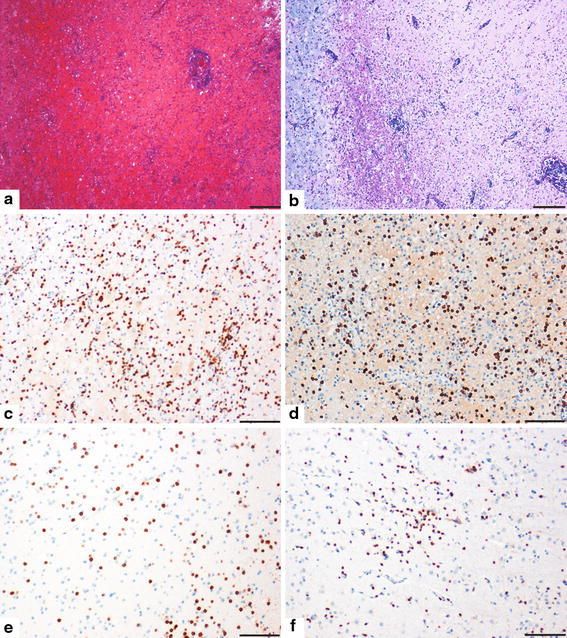
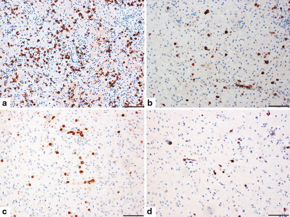
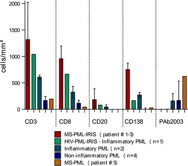
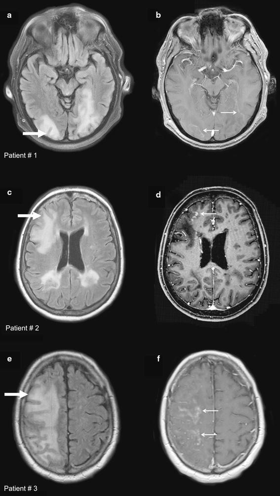
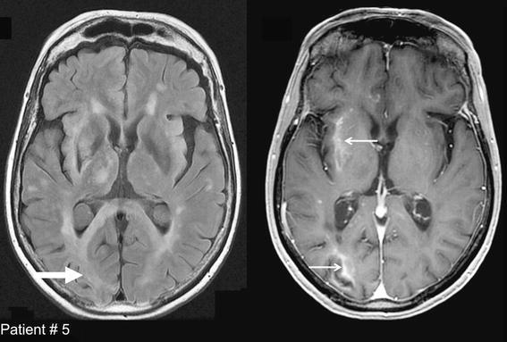
Natalizumab is an approved medication for highly active multiple sclerosis (MS). Progressive multifocal leukoencephalopathy (PML) may occur as a severe side effect of this drug. Here, we describe pathological and radiological characteristics of immune reconstitution inflammatory syndrome (IRIS), which occurs in natalizumab-associated PML after the cessation of therapy, and we differentiate it from ongoing PML. Brain biopsy tissue and MRI scans from five MS patients with natalizumab-associated PML were analyzed and their histology compared with non-MS PML. Histology showed an extensive CD8-dominated T cell infiltrate and numerous macrophages within lesions, and in nondemyelinated white and grey matter, in four out of five cases. Few or no virally infected cells were found. This was indicative of IRIS as known from HIV patients with PML. Outstandingly high numbers of plasma cells were present as compared to non-MS PML and typical MS lesions. MRI was compatible with IRIS, revealing enlarging lesions with a band-like or speckled contrast enhancement either at the lesion edge or within lesions. Only the fifth patient showed typical PML pathology, with low inflammation and high numbers of virally infected cells. This patient showed a similar interval between drug withdrawal and biopsy (3.5 months) to the rest of the cohort (range 2.5-4 months). MRI could not differentiate between PML-associated IRIS and ongoing PML. We describe in detail the histopathology of IRIS in natalizumab-associated PML. PML-IRIS, ongoing PML infection, and MS exacerbation may be impossible to discern clinically alone. MRI may provide some clues for distinguishing different pathologies that can be differentiated histologically. In our individual cases, biopsy helped to clarify diagnoses in natalizumab-associated PML.
那他珠单抗是一种已被批准用于治疗高度活跃性多发性硬化症(MS)的药物。进行性多灶性白质脑病(PML)可能是这种药物的一种严重副作用。在此,我们描述了免疫重建炎症综合征(IRIS)的病理和影像学特征,该综合征发生在停止治疗后与那他珠单抗相关的 PML 中,我们将其与持续的 PML 区分开来。对 5 名患有那他珠单抗相关性 PML 的 MS 患者的脑活检组织和 MRI 扫描进行了分析,并将其组织学与非 MS PML 进行了比较。组织学显示,在 4 例中,病变内广泛存在 CD8 主导的 T 细胞浸润和大量巨噬细胞,在非脱髓鞘的白质和灰质中也是如此。很少或没有发现病毒感染的细胞。这表明存在 IRIS,这与 HIV 患者的 PML 相似。与非 MS PML 和典型的 MS 病变相比,浆细胞数量明显增多。MRI 与 IRIS 相符,显示出病变扩大,在病变边缘或病变内呈带状或斑点状对比增强。只有第 5 名患者显示出典型的 PML 病理学,炎症程度低,病毒感染细胞数量多。这名患者在停药和活检之间的间隔(3.5 个月)与队列中的其他患者(2.5-4 个月)相似。MRI 无法区分 PML 相关的 IRIS 和持续的 PML。我们详细描述了那他珠单抗相关性 PML 中 IRIS 的组织病理学。PML-IRIS、持续性 PML 感染和 MS 恶化在临床上可能无法单独区分。MRI 可能为区分不同的病理提供一些线索,这些病理可以通过组织学进行区分。在我们的个别病例中,活检有助于澄清那他珠单抗相关性 PML 的诊断。