• 文献检索
  • 文档翻译
  • 深度研究
  • 学术资讯
  • Suppr Zotero 插件Zotero 插件
  • 邀请有礼
  • 套餐&价格
  • 历史记录
应用&插件
Suppr Zotero 插件Zotero 插件浏览器插件Mac 客户端Windows 客户端微信小程序
定价
高级版会员购买积分包购买API积分包
服务
文献检索文档翻译深度研究API 文档MCP 服务
关于我们
关于 Suppr公司介绍联系我们用户协议隐私条款
关注我们

Suppr 超能文献

核心技术专利:CN118964589B侵权必究
粤ICP备2023148730 号-1Suppr @ 2026

文献检索

告别复杂PubMed语法,用中文像聊天一样搜索,搜遍4000万医学文献。AI智能推荐,让科研检索更轻松。

立即免费搜索

文件翻译

保留排版,准确专业,支持PDF/Word/PPT等文件格式,支持 12+语言互译。

免费翻译文档

深度研究

AI帮你快速写综述,25分钟生成高质量综述,智能提取关键信息,辅助科研写作。

立即免费体验

通过慢病毒介导的强直性肌营养不良1型(DM1)成纤维细胞的MyoD转导产生的成肌细胞可用于治疗分子的检测。

Myoblasts generated by lentiviral mediated MyoD transduction of myotonic dystrophy type 1 (DM1) fibroblasts can be used for assays of therapeutic molecules.

作者信息

Larsen Jan, Pettersson Olof J, Jakobsen Maria, Thomsen Rune, Pedersen Christina B, Hertz Jens M, Gregersen Niels, Corydon Thomas J, Jensen Thomas G

机构信息

Department of Biomedicine, Aarhus University, Aarhus, Denmark.

出版信息

BMC Res Notes. 2011 Nov 11;4:490. doi: 10.1186/1756-0500-4-490.

DOI:10.1186/1756-0500-4-490
PMID:22078098
原文链接:https://pmc.ncbi.nlm.nih.gov/articles/PMC3226528/
Abstract

BACKGROUND

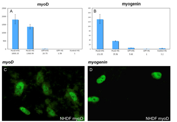
Myotonic dystrophy type 1 (DM1) is the most common muscle dystrophy in adults. The disease is caused by a triplet expansion in the 3'end of the myotonic dystrophy protein kinase (DMPK) gene. In order to develop a human cell model for investigation of possible effects of antisense and RNAi effector molecules we have used lentiviral mediated myoD-forced myogenesis of DM1 patient fibroblasts.

FINDINGS

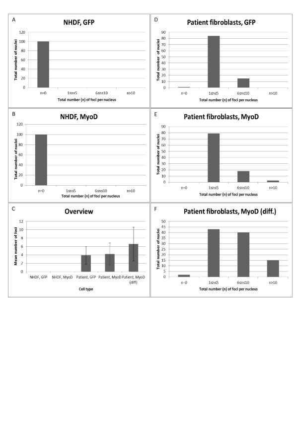
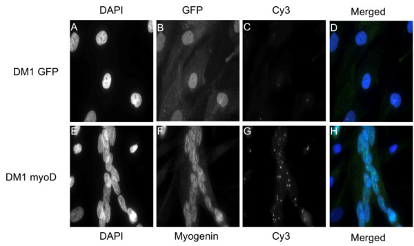
Transduced fibroblasts show a multinuclear phenotype and express the differentiation marker myogenin. Furthermore, fluorescence in situ hybridization (FISH) analysis revealed a statistical significant increase in the amount of nuclear foci in DM1 patient fibroblasts after myogenesis. Finally, no nuclear foci were found after treatment with oligonucleotides targeting the repeat expansions.

CONCLUSIONS

The abundance of nuclear foci in DM1 patient fibroblasts increase following myogenesis, as visualized by FISH analysis. Foci were eradicated after treatment with antisense oligonucleotides. Thus, we propose that the current cell model is suitable for testing of novel treatment modalities.

摘要

背景

1型强直性肌营养不良(DM1)是成人中最常见的肌肉营养不良症。该疾病由强直性肌营养不良蛋白激酶(DMPK)基因3'端的三联体扩增引起。为了建立一个人类细胞模型来研究反义寡核苷酸和RNA干扰效应分子的潜在作用,我们利用慢病毒介导的DM1患者成纤维细胞的肌分化因子(myoD)诱导成肌作用。

研究结果

转导后的成纤维细胞呈现多核表型并表达分化标志物肌细胞生成素。此外,荧光原位杂交(FISH)分析显示,成肌作用后DM1患者成纤维细胞核灶数量有统计学意义的增加。最后,用靶向重复扩增序列的寡核苷酸处理后未发现核灶。

结论

FISH分析显示,DM1患者成纤维细胞成肌作用后核灶数量增加。反义寡核苷酸处理后核灶消失。因此,我们认为当前的细胞模型适用于测试新的治疗方法。

https://cdn.ncbi.nlm.nih.gov/pmc/blobs/26cd/3226528/4e70c591839f/1756-0500-4-490-5.jpg
https://cdn.ncbi.nlm.nih.gov/pmc/blobs/26cd/3226528/a8b13d2f8118/1756-0500-4-490-1.jpg
https://cdn.ncbi.nlm.nih.gov/pmc/blobs/26cd/3226528/84c90a971dd8/1756-0500-4-490-2.jpg
https://cdn.ncbi.nlm.nih.gov/pmc/blobs/26cd/3226528/97f8ec3b04d8/1756-0500-4-490-3.jpg
https://cdn.ncbi.nlm.nih.gov/pmc/blobs/26cd/3226528/31f11e3f6c44/1756-0500-4-490-4.jpg
https://cdn.ncbi.nlm.nih.gov/pmc/blobs/26cd/3226528/4e70c591839f/1756-0500-4-490-5.jpg

相似文献

1
Myoblasts generated by lentiviral mediated MyoD transduction of myotonic dystrophy type 1 (DM1) fibroblasts can be used for assays of therapeutic molecules.通过慢病毒介导的强直性肌营养不良1型(DM1)成纤维细胞的MyoD转导产生的成肌细胞可用于治疗分子的检测。
BMC Res Notes. 2011 Nov 11;4:490. doi: 10.1186/1756-0500-4-490.
2
Fuchs' Endothelial Corneal Dystrophy and RNA Foci in Patients With Myotonic Dystrophy.强直性肌营养不良患者的富克斯角膜内皮营养不良与RNA病灶
Invest Ophthalmol Vis Sci. 2017 Sep 1;58(11):4579-4585. doi: 10.1167/iovs.17-22350.
3
Immortalized human myotonic dystrophy muscle cell lines to assess therapeutic compounds.永生化人类强直性肌营养不良症肌肉细胞系用于评估治疗性化合物。
Dis Model Mech. 2017 Apr 1;10(4):487-497. doi: 10.1242/dmm.027367. Epub 2017 Feb 10.
4
Mutant DMPK 3'-UTR transcripts disrupt C2C12 myogenic differentiation by compromising MyoD.突变型DMPK 3'-UTR转录本通过损害MyoD来破坏C2C12成肌分化。
J Cell Biol. 2002 Nov 11;159(3):419-29. doi: 10.1083/jcb.200206020.
5
Recapitulating muscle disease phenotypes with myotonic dystrophy 1 induced pluripotent stem cells: a tool for disease modeling and drug discovery.利用肌强直性营养不良 1 诱导的多能干细胞再现肌肉疾病表型:疾病建模和药物发现的工具。
Dis Model Mech. 2018 Jul 18;11(7):dmm034728. doi: 10.1242/dmm.034728.
6
Hammerhead ribozyme-mediated destruction of nuclear foci in myotonic dystrophy myoblasts.锤头状核酶介导的强直性肌营养不良成肌细胞中核灶的破坏。
Mol Ther. 2003 May;7(5 Pt 1):670-80. doi: 10.1016/s1525-0016(03)00068-6.
7
Decreased levels of myotonic dystrophy protein kinase (DMPK) and delayed differentiation in human myotonic dystrophy myoblasts.强直性肌营养不良蛋白激酶(DMPK)水平降低与人类强直性肌营养不良成肌细胞分化延迟。
Neuromuscul Disord. 2001 Nov;11(8):728-35. doi: 10.1016/s0960-8966(01)00226-7.
8
Sense and Antisense DMPK RNA Foci Accumulate in DM1 Tissues during Development.在发育过程中,有义链和反义链 DMPK RNA 病灶在 DM1 组织中积累。
PLoS One. 2015 Sep 4;10(9):e0137620. doi: 10.1371/journal.pone.0137620. eCollection 2015.
9
Muscleblind localizes to nuclear foci of aberrant RNA in myotonic dystrophy types 1 and 2.在1型和2型强直性肌营养不良中,肌盲蛋白定位于异常RNA的核灶。
Hum Mol Genet. 2001 Sep 15;10(19):2165-70. doi: 10.1093/hmg/10.19.2165.
10
Promising AAV.U7snRNAs vectors targeting improve DM1 hallmarks in patient-derived cell lines.靶向改善患者来源细胞系中DM1特征的有前景的AAV.U7snRNAs载体。
Front Cell Dev Biol. 2023 Jun 15;11:1181040. doi: 10.3389/fcell.2023.1181040. eCollection 2023.

引用本文的文献

1
Repeat length as a key determinant for disease severity and antisense oligonucleotide activity in myotonic dystrophy type 1.重复序列长度是1型强直性肌营养不良症疾病严重程度和反义寡核苷酸活性的关键决定因素。
Mol Ther Methods Clin Dev. 2025 Jun 2;33(3):101502. doi: 10.1016/j.omtm.2025.101502. eCollection 2025 Sep 11.
2
Efficacy of Cystic Fibrosis Transmembrane Regulator Corrector C17 in Beta-Sarcoglycanopathy-Assessment of Patient's Primary Myotubes.囊性纤维化跨膜调节因子校正剂C17在β-肌聚糖病中的疗效——患者原代肌管评估
Int J Mol Sci. 2024 Dec 11;25(24):13313. doi: 10.3390/ijms252413313.
3
Promising AAV.U7snRNAs vectors targeting improve DM1 hallmarks in patient-derived cell lines.

本文引用的文献

1
HTS-Compatible Patient-Derived Cell-Based Assay to Identify Small Molecule Modulators of Aberrant Splicing in Myotonic Dystrophy Type 1.用于鉴定1型强直性肌营养不良中异常剪接小分子调节剂的与高通量筛选兼容的患者来源细胞检测法
Curr Chem Genomics. 2010 Mar 19;4:9-18. doi: 10.2174/1875397301004010009.
2
Normal myogenesis and increased apoptosis in myotonic dystrophy type-1 muscle cells.正常的成肌发生和肌强直性营养不良 1 型肌肉细胞中的凋亡增加。
Cell Death Differ. 2010 Aug;17(8):1315-24. doi: 10.1038/cdd.2010.33. Epub 2010 Apr 30.
3
Aberrant alternative splicing and extracellular matrix gene expression in mouse models of myotonic dystrophy.
靶向改善患者来源细胞系中DM1特征的有前景的AAV.U7snRNAs载体。
Front Cell Dev Biol. 2023 Jun 15;11:1181040. doi: 10.3389/fcell.2023.1181040. eCollection 2023.
4
Pluripotent Stem Cells in Disease Modeling and Drug Discovery for Myotonic Dystrophy Type 1.多能干细胞在肌强直性营养不良 1 型疾病建模和药物发现中的应用。
Cells. 2023 Feb 10;12(4):571. doi: 10.3390/cells12040571.
5
Loss of the Nuclear Envelope Protein LAP1B Disrupts the Myogenic Differentiation of Patient-Derived Fibroblasts.核包膜蛋白 LAP1B 的缺失破坏了患者来源的成纤维细胞的肌生成分化。
Int J Mol Sci. 2022 Nov 6;23(21):13615. doi: 10.3390/ijms232113615.
6
Recent Progress and Challenges in the Development of Antisense Therapies for Myotonic Dystrophy Type 1.1 型肌强直性营养不良反义疗法的研究进展与挑战
Int J Mol Sci. 2022 Nov 1;23(21):13359. doi: 10.3390/ijms232113359.
7
Brain Pathogenesis and Potential Therapeutic Strategies in Myotonic Dystrophy Type 1.1型强直性肌营养不良症的脑发病机制及潜在治疗策略
Front Aging Neurosci. 2021 Nov 15;13:755392. doi: 10.3389/fnagi.2021.755392. eCollection 2021.
8
Recovery in the Myogenic Program of Congenital Myotonic Dystrophy Myoblasts after Excision of the Expanded (CTG) Repeat.先天性肌强直性营养不良成肌细胞中扩张(CTG)重复序列切除后的成肌细胞再生。
Int J Mol Sci. 2019 Nov 13;20(22):5685. doi: 10.3390/ijms20225685.
9
Intrinsic Myogenic Potential of Skeletal Muscle-Derived Pericytes from Patients with Myotonic Dystrophy Type 1.1型强直性肌营养不良症患者骨骼肌来源周细胞的内在肌源性潜能
Mol Ther Methods Clin Dev. 2019 Sep 12;15:120-132. doi: 10.1016/j.omtm.2019.09.002. eCollection 2019 Dec 13.
10
Distinct pathological signatures in human cellular models of myotonic dystrophy subtypes.不同肌强直性营养不良亚型的人类细胞模型中的独特病理特征。
JCI Insight. 2019 Mar 21;4(6). doi: 10.1172/jci.insight.122686.
肌强直性营养不良症小鼠模型中的异常选择性剪接和细胞外基质基因表达。
Nat Struct Mol Biol. 2010 Feb;17(2):187-93. doi: 10.1038/nsmb.1720. Epub 2010 Jan 24.
4
DNA/polyethyleneimine/hyaluronic acid small complex particles and tumor suppression in mice.DNA/聚乙烯亚胺/透明质酸小复合粒子对小鼠的肿瘤抑制作用。
Biomaterials. 2010 Apr;31(10):2912-8. doi: 10.1016/j.biomaterials.2009.12.032. Epub 2010 Jan 4.
5
Pathogenic mechanisms of myotonic dystrophy.肌强直性营养不良的发病机制。
Biochem Soc Trans. 2009 Dec;37(Pt 6):1281-6. doi: 10.1042/BST0371281.
6
Pentamidine reverses the splicing defects associated with myotonic dystrophy.喷他脒可逆转与强直性肌营养不良相关的剪接缺陷。
Proc Natl Acad Sci U S A. 2009 Nov 3;106(44):18551-6. doi: 10.1073/pnas.0903234106. Epub 2009 Oct 12.
7
Triplet-repeat oligonucleotide-mediated reversal of RNA toxicity in myotonic dystrophy.三联体重复寡核苷酸介导的强直性肌营养不良症中RNA毒性的逆转
Proc Natl Acad Sci U S A. 2009 Aug 18;106(33):13915-20. doi: 10.1073/pnas.0905780106. Epub 2009 Aug 10.
8
Reversal of RNA dominance by displacement of protein sequestered on triplet repeat RNA.通过置换隔离于三联体重复RNA上的蛋白质来逆转RNA主导地位。
Science. 2009 Jul 17;325(5938):336-9. doi: 10.1126/science.1173110.
9
Amelioration of psoriasis by anti-TNF-alpha RNAi in the xenograft transplantation model.在异种移植移植模型中通过抗 TNF-α RNAi 改善银屑病。
Mol Ther. 2009 Oct;17(10):1743-53. doi: 10.1038/mt.2009.141. Epub 2009 Jun 30.
10
Immortalized skin fibroblasts expressing conditional MyoD as a renewable and reliable source of converted human muscle cells to assess therapeutic strategies for muscular dystrophies: validation of an exon-skipping approach to restore dystrophin in Duchenne muscular dystrophy cells.表达条件性 MyoD 的永生化皮肤成纤维细胞作为可转化的人类肌肉细胞的可再生和可靠来源,用于评估肌肉疾病的治疗策略:exon-skipping 方法在恢复杜氏肌营养不良症细胞中的肌营养不良蛋白的验证。
Hum Gene Ther. 2009 Jul;20(7):784-90. doi: 10.1089/hum.2008.163.