Structural and Computational Biology Unit, European Molecular Biology Laboratory, Heidelberg, Germany.
PLoS Biol. 2011 Nov;9(11):e1001196. doi: 10.1371/journal.pbio.1001196. Epub 2011 Nov 15.
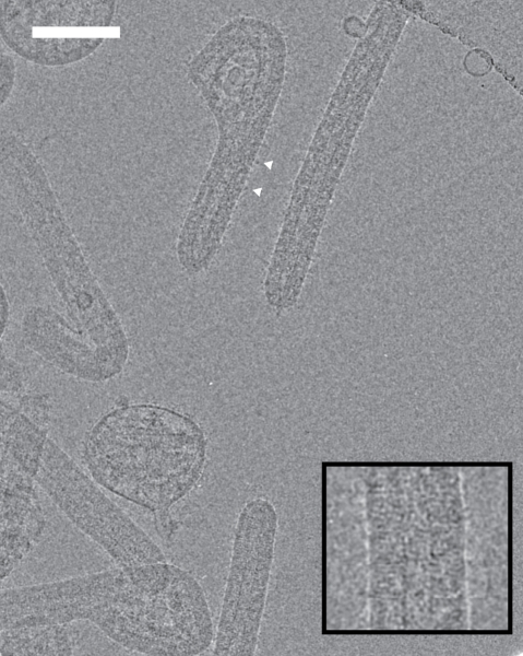
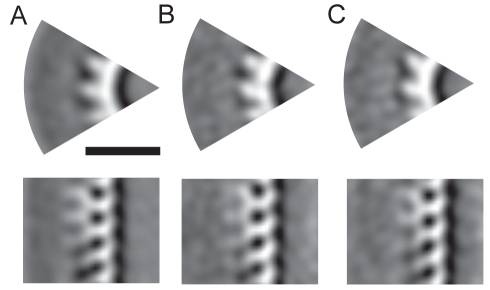
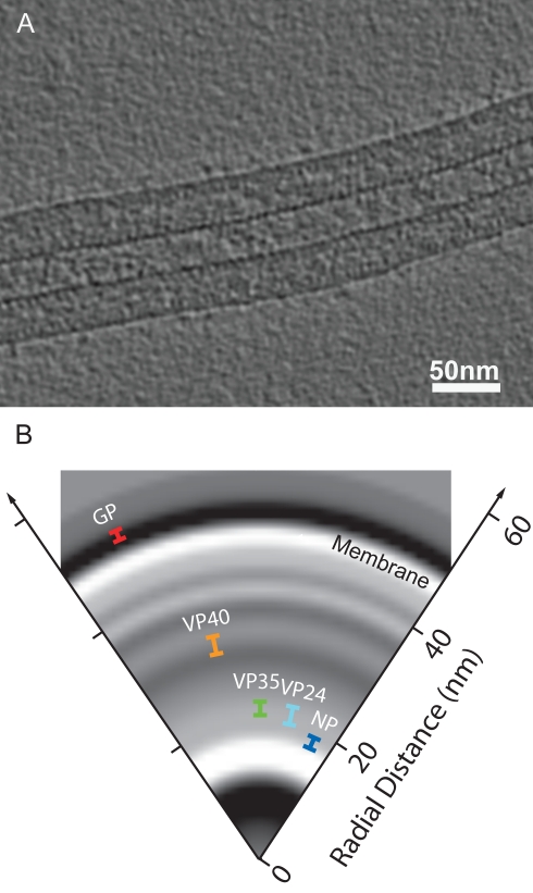
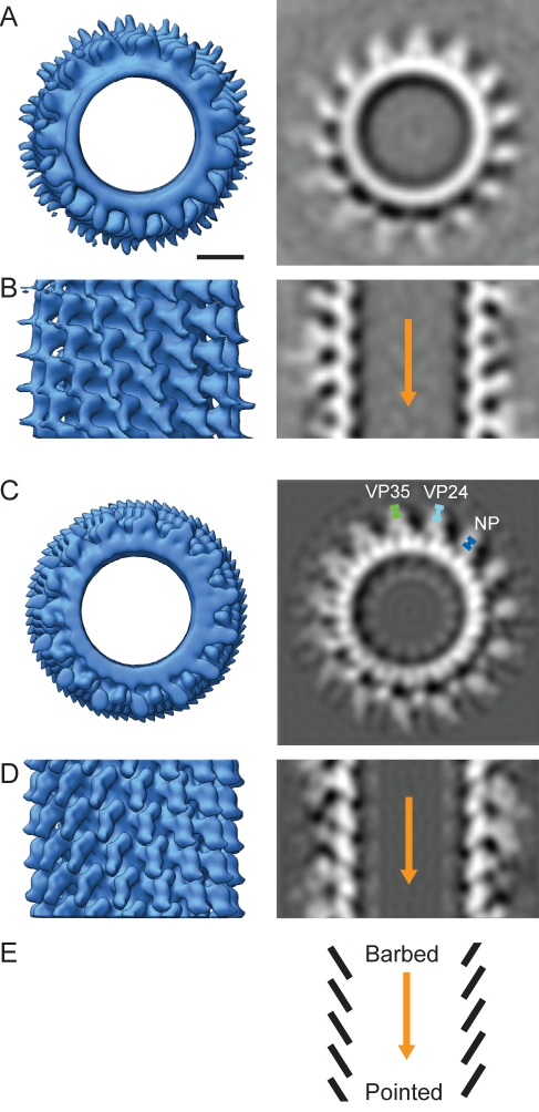
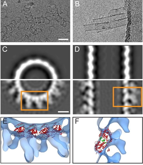
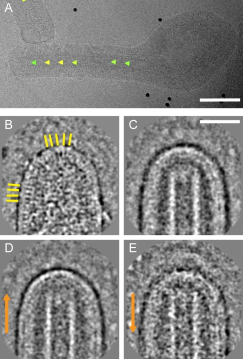
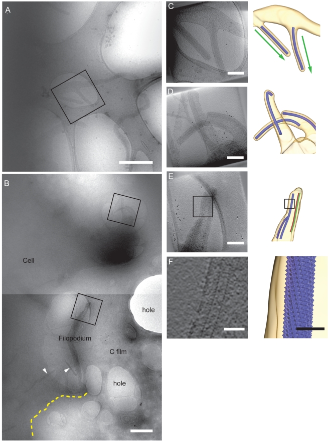
Several major human pathogens, including the filoviruses, paramyxoviruses, and rhabdoviruses, package their single-stranded RNA genomes within helical nucleocapsids, which bud through the plasma membrane of the infected cell to release enveloped virions. The virions are often heterogeneous in shape, which makes it difficult to study their structure and assembly mechanisms. We have applied cryo-electron tomography and sub-tomogram averaging methods to derive structures of Marburg virus, a highly pathogenic filovirus, both after release and during assembly within infected cells. The data demonstrate the potential of cryo-electron tomography methods to derive detailed structural information for intermediate steps in biological pathways within intact cells. We describe the location and arrangement of the viral proteins within the virion. We show that the N-terminal domain of the nucleoprotein contains the minimal assembly determinants for a helical nucleocapsid with variable number of proteins per turn. Lobes protruding from alternate interfaces between each nucleoprotein are formed by the C-terminal domain of the nucleoprotein, together with viral proteins VP24 and VP35. Each nucleoprotein packages six RNA bases. The nucleocapsid interacts in an unusual, flexible "Velcro-like" manner with the viral matrix protein VP40. Determination of the structures of assembly intermediates showed that the nucleocapsid has a defined orientation during transport and budding. Together the data show striking architectural homology between the nucleocapsid helix of rhabdoviruses and filoviruses, but unexpected, fundamental differences in the mechanisms by which the nucleocapsids are then assembled together with matrix proteins and initiate membrane envelopment to release infectious virions, suggesting that the viruses have evolved different solutions to these conserved assembly steps.
几种主要的人类病原体,包括丝状病毒、副粘病毒和弹状病毒,将其单链 RNA 基因组包装在螺旋核衣壳内,核衣壳通过感染细胞的质膜出芽释放包膜病毒。病毒的形状通常是异质的,这使得研究其结构和组装机制变得困难。我们已经应用冷冻电子断层扫描和亚断层平均方法来获得马尔堡病毒(一种高致病性丝状病毒)的结构,这些结构既有在释放后的,也有在感染细胞内组装过程中的。这些数据表明,冷冻电子断层扫描方法有可能为完整细胞内生物途径的中间步骤提供详细的结构信息。我们描述了病毒蛋白在病毒粒子中的位置和排列。我们表明,核蛋白的 N 端结构域包含形成具有可变蛋白数每圈的螺旋核衣壳的最小组装决定因素。每个核蛋白之间的交替界面突出的叶瓣由核蛋白的 C 端结构域与病毒蛋白 VP24 和 VP35 共同形成。每个核蛋白包装六个 RNA 碱基。核衣壳以一种不寻常的、灵活的“魔术贴样”方式与病毒基质蛋白 VP40 相互作用。组装中间体结构的测定表明,核衣壳在运输和出芽过程中有一个确定的取向。这些数据表明,弹状病毒和丝状病毒的核衣壳螺旋具有惊人的结构同源性,但在核衣壳与基质蛋白组装并启动膜包裹以释放感染性病毒颗粒的机制方面存在出乎意料的根本差异,这表明病毒已经进化出不同的解决方案来应对这些保守的组装步骤。