Department of Pathology, University of Aberdeen, Aberdeen, United Kingdom.
PLoS One. 2011;6(11):e27718. doi: 10.1371/journal.pone.0027718. Epub 2011 Nov 18.
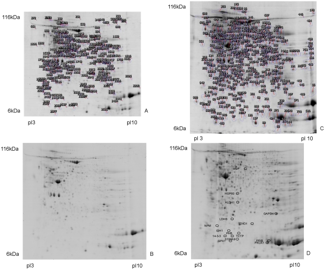
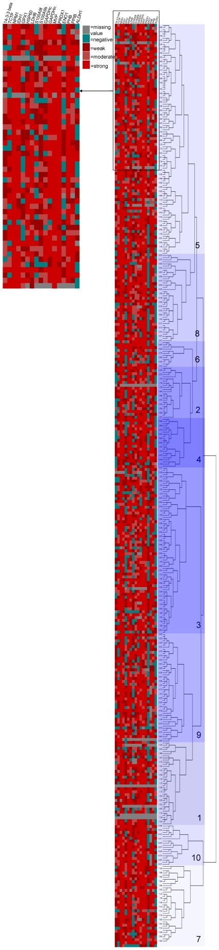
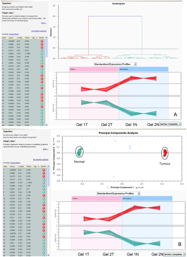
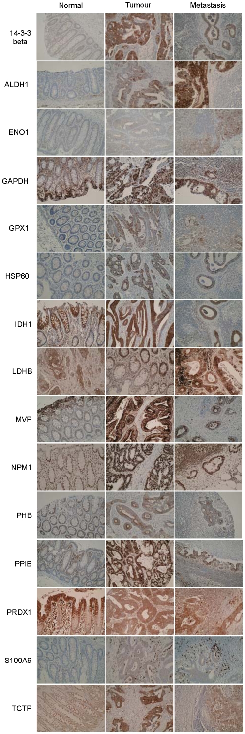
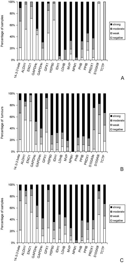
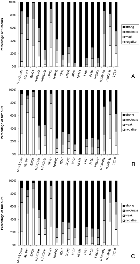
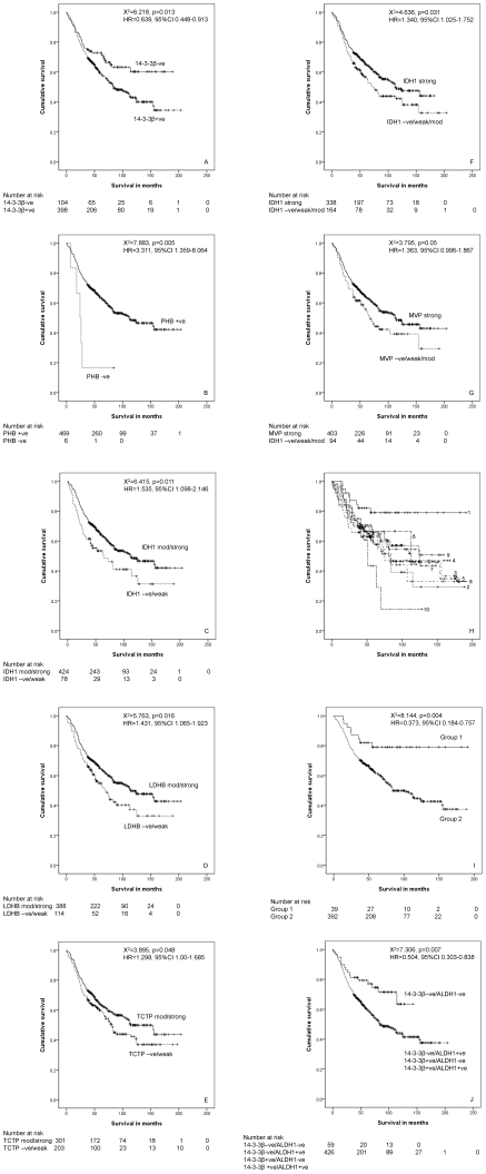
Colorectal cancer is one of the commonest types of cancer and there is requirement for the identification of prognostic biomarkers. In this study protein expression profiles have been established for colorectal cancer and normal colonic mucosa by proteomics using a combination of two dimensional gel electrophoresis with fresh frozen sections of paired Dukes B colorectal cancer and normal colorectal mucosa (n = 28), gel image analysis and high performance liquid chromatography-tandem mass spectrometry. Hierarchical cluster analysis and principal components analysis showed that the protein expression profiles of colorectal cancer and normal colonic mucosa clustered into distinct patterns of protein expression. Forty-five proteins were identified as showing at least 1.5 times increased expression in colorectal cancer and the identity of these proteins was confirmed by liquid chromatography-tandem mass spectrometry. Fifteen proteins that showed increased expression were validated by immunohistochemistry using a well characterised colorectal cancer tissue microarray containing 515 primary colorectal cancer, 224 lymph node metastasis and 50 normal colonic mucosal samples. The proteins that showed the greatest degree of overexpression in primary colorectal cancer compared with normal colonic mucosa were heat shock protein 60 (p<0.001), S100A9 (p<0.001) and translationally controlled tumour protein (p<0.001). Analysis of proteins individually identified 14-3-3β as a prognostic biomarker (χ² = 6.218, p = 0.013, HR = 0.639, 95%CI 0.448-0.913). Hierarchical cluster analysis identified distinct phenotypes associated with survival and a two-protein signature consisting of 14-3-3β and aldehyde dehydrogenase 1 was identified as showing prognostic significance (χ² = 7.306, p = 0.007, HR = 0.504, 95%CI 0.303-0.838) and that remained independently prognostic (p = 0.01, HR = 0.416, 95%CI 0.208-0.829) in a multivariate model.
结直肠癌是最常见的癌症类型之一,因此需要鉴定其预后生物标志物。本研究采用二维凝胶电泳联合新鲜冷冻结直肠肿瘤及正常黏膜配对组织(n=28)的图像分析和高效液相色谱-串联质谱技术建立了结直肠癌和正常结直肠黏膜的蛋白质表达谱。聚类分析和主成分分析显示,结直肠癌和正常结直肠黏膜的蛋白质表达谱聚类为明显不同的蛋白质表达模式。有 45 种蛋白质在结直肠癌中的表达至少增加了 1.5 倍,通过液相色谱-串联质谱技术对这些蛋白质的身份进行了确认。通过使用包含 515 例原发性结直肠癌、224 例淋巴结转移和 50 例正常结直肠黏膜样本的经充分鉴定的结直肠癌组织微阵列进行免疫组织化学验证,验证了 15 种表达增加的蛋白质。与正常结直肠黏膜相比,在原发性结直肠癌中表达增加最多的蛋白质为热休克蛋白 60(p<0.001)、S100A9(p<0.001)和翻译控制肿瘤蛋白(p<0.001)。对蛋白质的分析单独鉴定出 14-3-3β 为预后生物标志物(χ²=6.218,p=0.013,HR=0.639,95%CI 0.448-0.913)。聚类分析确定了与生存相关的不同表型,并且发现由 14-3-3β 和醛脱氢酶 1 组成的双蛋白特征具有预后意义(χ²=7.306,p=0.007,HR=0.504,95%CI 0.303-0.838),并且在多变量模型中仍然具有独立的预后意义(p=0.01,HR=0.416,95%CI 0.208-0.829)。