Department of Psychiatry and Clinical Neuroscience, Osaka University Graduate School of Medicine, Suita City, Osaka, Japan.
PLoS One. 2011;6(11):e27863. doi: 10.1371/journal.pone.0027863. Epub 2011 Nov 18.
It is unclear whether, like in schizophrenia, psychosis-related disruption in connectivity between certain regions, as an index of intrinsic functional disintegration, occurs in schizophrenia-like psychosis of epilepsy (SLPE). In this study, we sought to determine abnormal patterns of resting-state EEG oscillations and functional connectivity in patients with SLPE, compared with nonpsychotic epilepsy patients, and to assess correlations with psychopathological deficits.
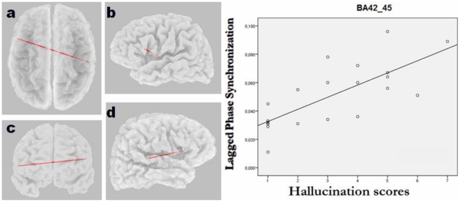
METHODOLOGY/PRINCIPAL FINDINGS: Resting EEG was recorded in 21 patients with focal epilepsy and SLPE and in 21 clinically-matched non-psychotic epilepsy controls. Source current density and functional connectivity were determined using eLORETA software. For connectivity analysis, a novel nonlinear connectivity measure called "lagged phase synchronization" was used. We found increased theta oscillations in regions involved in the default mode network (DMN), namely the medial and lateral parietal cortex bilaterally in the psychotic patients relative to their nonpsychotic counterparts. In addition, patients with psychosis had increased beta temporo-prefrontal connectivity in the hemisphere with predominant seizure focus. This functional connectivity in temporo-prefrontal circuits correlated with positive symptoms. Additionally, there was increased interhemispheric phase synchronization between the auditory cortex of the affected temporal lobe and the Broca's area correlating with auditory hallucination scores.
CONCLUSIONS/SIGNIFICANCE: In addition to dysfunction of parietal regions that are part of the DMN, resting-state disrupted connectivity of the medial temporal cortex with prefrontal areas that are either involved in the DMN or implicated in psychopathological dysfunction may be critical to schizophrenia-like psychosis, especially in individuals with temporal lobe epilepsy. This suggests that DMN deficits might be a core neurobiological feature of the disorder, and that abnormalities in theta oscillations and beta phase synchronization represent the underlying neural activity.
在癫痫相关的精神分裂样精神病(SLPE)中,是否存在特定区域之间的连接中断,作为内在功能分离的指标,类似于精神分裂症,目前尚不清楚。在这项研究中,我们试图确定与非精神病性癫痫患者相比,SLPE 患者静息状态 EEG 振荡和功能连接的异常模式,并评估其与精神病理学缺陷的相关性。
方法/主要发现:记录了 21 例局灶性癫痫和 SLPE 患者以及 21 例临床匹配的非精神病性癫痫对照患者的静息 EEG。使用 eLORETA 软件确定源电流密度和功能连接。对于连接分析,使用一种称为“滞后相位同步”的新型非线性连接测量方法。我们发现,与非精神病性患者相比,精神病患者的默认模式网络(DMN)相关区域(双侧内侧和外侧顶叶皮层)中的θ振荡增加。此外,在以优势癫痫灶为主的半球中,精神病患者的颞叶-前额叶功能连接增加。颞叶-前额叶回路的这种功能连接与阳性症状相关。此外,受影响的颞叶听觉皮层与布罗卡区之间的半球间相位同步增加,与幻听评分相关。
结论/意义:除了 DMN 部分的顶叶区域功能障碍外,内侧颞叶与前额叶区域的静息状态连接中断,这些区域要么参与 DMN,要么与精神病理学功能障碍有关,这可能对精神分裂样精神病至关重要,尤其是在颞叶癫痫患者中。这表明 DMN 缺陷可能是该疾病的核心神经生物学特征,而θ振荡和β相位同步异常代表潜在的神经活动。