Yang Xuejiao, Liu Bingqian, Bai Yujing, Chen Min, Li Yiqing, Chen Mengfei, Wei Yantao, Ge Jian, Zhuo Yehong
State Key Laboratory of Ophthalmology, Zhongshan Ophthalmic Center, Sun Yat-sen University, Guangzhou, China.
Mol Vis. 2011;17:2978-85. Epub 2011 Nov 16.
To investigate the effect of elevated hydrostatic pressure on the expression and distribution of zonula occludens-1 (ZO-1), and its effect on cytoskeleton and focal adhesion in immortal human trabecular meshwork cells (iHTM) and glaucomatous human trabecular meshwork cells (GTM(3)).
iHTM and GTM(3) were exposed to 60 mmHg hydrostatic pressure for 6, 12, and 24 h. As a control, the cells were incubated simultaneously in a conventional incubator. Morphology changes were observed with an inverted microscope. The expression of ZO-1was examined with western blot, and the distribution of ZO-1 was assessed by immunofluorescence. Actin cytoskeleton and focal adhesion (vinculin) were also assessed by immunofluorescence. Data were analyzed with commercial data analysis software and a p<0.05 was considered to be statistically significant.
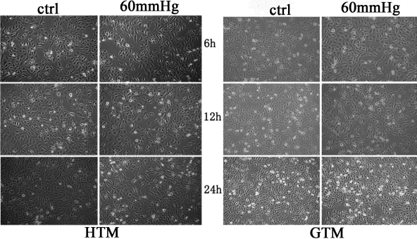
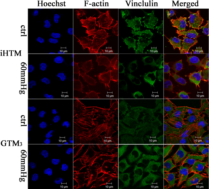
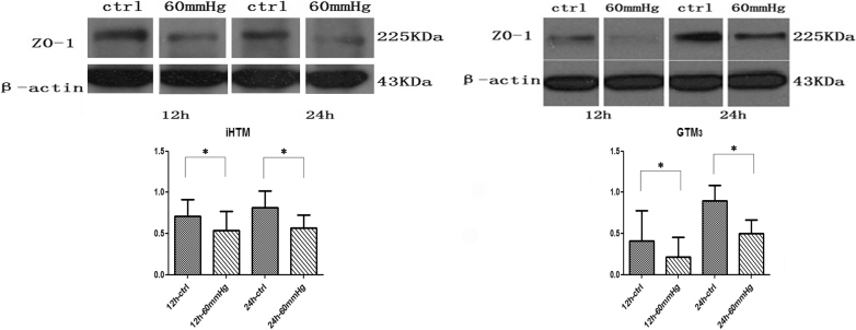
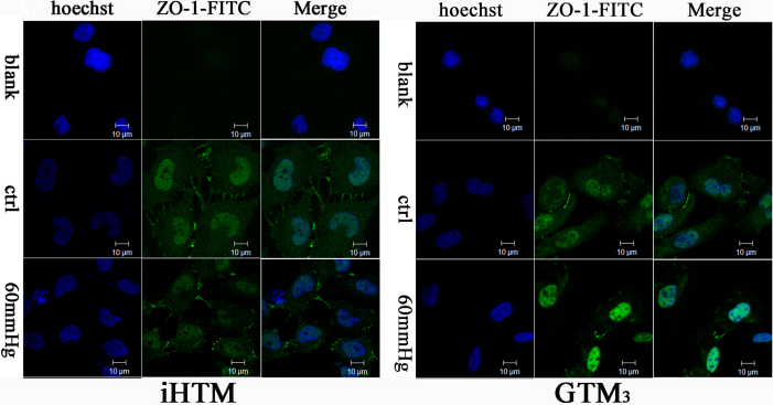
There was no evident morphology change after 24 h culture in 60 mmHg pressure in iHTM and GTM(3). However, in both iHTM and GTM(3), elevated pressure attenuated the expression of ZO-1 at 12 h and 24 h, detected by western blot. Meanwhile, high pressure disrupted the organization of ZO-1, actin cytoskeleton, and vinculin, assessed by immunofluorescence. When comparing iHTM with GTM(3), the distribution of ZO-1 and vinculin in GTM(3) was not as regular as that in iHTM. After exposuring in elevated pressure, the changes in GTM(3) were more obvious than that in iHTM.
Sustained pressure elevation may directly damage trabecular meshwork cells by injuring ZO-1, cytoskeleton, and foal adhesions. And GTM(3) was more susceptible to damage than iHTM. We suggest that elevated pressure seems to be not only the results of damaged TM, but also an important factor for the injury of TM cells, stop or reverse the process may help developing new target for the treatment of primary open angle glaucoma (POAG).
研究升高的静水压力对紧密连接蛋白-1(ZO-1)表达和分布的影响,及其对永生化人小梁网细胞(iHTM)和青光眼患者小梁网细胞(GTM(3))细胞骨架和粘着斑的影响。
将iHTM和GTM(3)暴露于60 mmHg静水压力下6、12和24小时。作为对照,细胞同时在传统培养箱中培养。用倒置显微镜观察形态变化。用蛋白质免疫印迹法检测ZO-1的表达,用免疫荧光法评估ZO-1的分布。肌动蛋白细胞骨架和粘着斑(纽蛋白)也通过免疫荧光进行评估。数据用商业数据分析软件进行分析,p<0.05被认为具有统计学意义。
在60 mmHg压力下培养24小时后,iHTM和GTM(3)均未出现明显的形态变化。然而,蛋白质免疫印迹法检测显示,在iHTM和GTM(3)中,升高的压力在12小时和24小时时均减弱了ZO-1的表达。同时,免疫荧光评估显示,高压破坏了ZO-1、肌动蛋白细胞骨架和纽蛋白的组织。当比较iHTM和GTM(3)时,GTM(3)中ZO-1和纽蛋白的分布不如iHTM规则。在暴露于升高的压力后,GTM(3)中的变化比iHTM更明显。
持续的压力升高可能通过损伤ZO-1、细胞骨架和粘着斑直接损害小梁网细胞。并且GTM(3)比iHTM更容易受到损伤。我们认为升高的压力似乎不仅是小梁网受损的结果,也是小梁网细胞损伤的重要因素,阻止或逆转这一过程可能有助于开发原发性开角型青光眼(POAG)治疗的新靶点。