Department of Anatomy and Cell Biology, and Kansas Intellectual and Developmental Disabilities Research Center, University of Kansas Medical School, 3901 Rainbow Blvd., MS 3051, HLSIC Rm. 2073, Kansas City, KS 66160, USA.
Mol Neurobiol. 2012 Feb;45(1):1-16. doi: 10.1007/s12035-011-8216-y. Epub 2011 Dec 2.
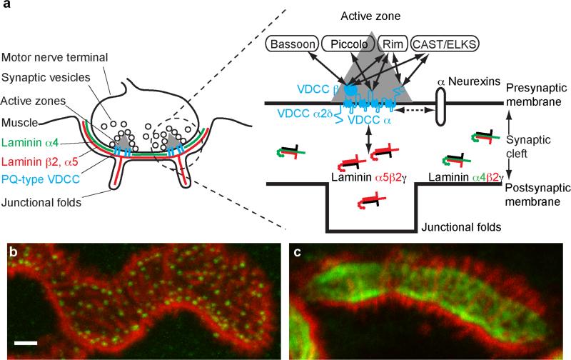
Organization of presynaptic active zones is essential for development, plasticity, and pathology of the nervous system. Recent studies indicate a trans-synaptic molecular mechanism that organizes the active zones by connecting the pre- and the postsynaptic specialization. The presynaptic component of this trans-synaptic mechanism is comprised of cytosolic active zone proteins bound to the cytosolic domains of voltage-dependent calcium channels (P/Q-, N-, and L-type) on the presynaptic membrane. The postsynaptic component of this mechanism is the synapse organizer (laminin β2) that is expressed by the postsynaptic cell and accumulates specifically on top of the postsynaptic specialization. The pre- and the postsynaptic components interact directly between the extracellular domains of calcium channels and laminin β2 to anchor the presynaptic protein complex in front of the postsynaptic specialization. Hence, the presynaptic calcium channel functions as a scaffolding protein for active zone organization and as an ion-conducting channel for synaptic transmission. In contrast to the requirement of calcium influx for synaptic transmission, the formation of the active zone does not require the calcium influx through the calcium channels. Importantly, the active zones of adult synapses are not stable structures and require maintenance for their integrity. Furthermore, aging or diseases of the central and peripheral nervous system impair the active zones. This review will focus on the molecular mechanisms that organize the presynaptic active zones and summarize recent findings at the neuromuscular junctions and other synapses.
突触前活性区的组织对于神经系统的发育、可塑性和病理学至关重要。最近的研究表明了一种跨突触的分子机制,通过连接突触前和突触后特化来组织活性区。这种跨突触机制的突触前成分包括与突触前膜上电压依赖性钙通道(P/Q-、N-和 L-型)胞质域结合的胞质活性区蛋白。该机制的突触后成分是突触组织者(层粘连蛋白β2),它由突触后细胞表达,并特异性地积累在突触后特化的顶部。该机制的前突触和后突触成分在钙通道和层粘连蛋白β2的细胞外结构域之间直接相互作用,将突触前蛋白复合物锚定在突触后特化的前面。因此,突触前钙通道作为活性区组织的支架蛋白和作为突触传递的离子通道发挥作用。与钙流入对于突触传递的要求相反,活性区的形成不需要钙通道通过钙流入。重要的是,成年突触的活性区不是稳定的结构,需要维持其完整性。此外,中枢和周围神经系统的衰老或疾病会损害活性区。这篇综述将重点介绍组织突触前活性区的分子机制,并总结神经肌肉接头和其他突触的最新发现。