Department of Urology, Hannover Medical School, (Carl-Neuberg-Strasse 1), Hannover, (30625), Germany.
BMC Urol. 2011 Dec 7;11:25. doi: 10.1186/1471-2490-11-25.
Caveolae play a significant role in disease phenotypes such as cancer, diabetes, bladder dysfunction, and muscular dystrophy. The aim of this study was to elucidate the caveolin-1 (CAV1) protein expression in renal cell cancer (RCC) and to determine its potential prognostic relevance.
289 clear cell RCC tissue specimens were collected from patients undergoing surgery for renal tumors. Both cytoplasmic and membranous CAV1 expression were determined by immunohistochemistry and correlated with clinical variables. Survival analysis was carried out for 169 evaluable patients with a median follow up of 80.5 months (interquartile range (IQR), 24.5-131.7 months).
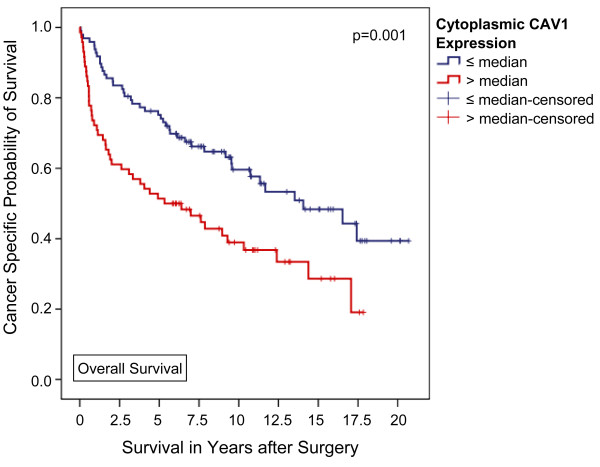
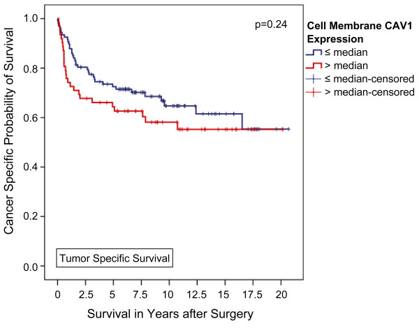
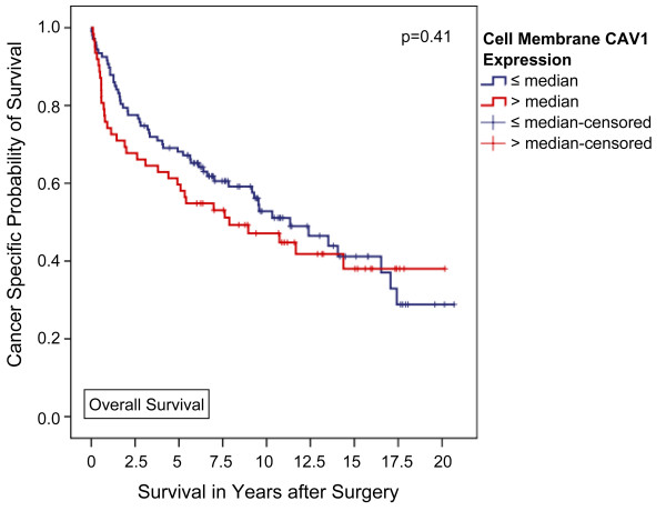
A high CAV1 expression in the tumor cell cytoplasm was significantly associated with male sex (p = 0.04), a positive nodal status (p = 0.04), and poor tumor differentiation (p = 0.04). In contrast, a higher than average (i.e. > median) CAV1 expression in tumor cell membranes was only linked to male sex (p = 0.03). Kaplan-Meier analysis disclosed significant differences in 5-year overall (51.4 vs. 75.2%, p = 0.001) and tumor specific survival (55.3 vs. 80.1%, p = 0.001) for patients with higher and lower than average cytoplasmic CAV1 expression levels, respectively. Applying multivariable Cox regression analysis a high CAV1 protein expression level in the tumor cell cytoplasm could be identified as an independent poor prognostic marker of both overall (p = 0.02) and tumor specific survival (p = 0.03) in clear cell RCC patients.
Over expression of caveolin-1 in the tumour cell cytoplasm predicts a poor prognosis of patients with clear cell RCC. CAV1 is likely to be a useful prognostic marker and may play an important role in tumour progression. Therefore, our data encourage further investigations to enlighten the role of CAV1 and its function as diagnostic and prognostic marker in serum and/or urine of RCC patients.
小窝在疾病表型中发挥着重要作用,如癌症、糖尿病、膀胱功能障碍和肌肉营养不良。本研究的目的是阐明小窝蛋白-1(CAV1)在肾细胞癌(RCC)中的蛋白表达,并确定其潜在的预后相关性。
收集 289 例接受肾肿瘤手术的透明细胞 RCC 组织标本。通过免疫组织化学法检测细胞质和膜小窝蛋白-1的表达,并与临床变量相关联。对 169 例可评估患者进行生存分析,中位随访时间为 80.5 个月(四分位间距(IQR),24.5-131.7 个月)。
肿瘤细胞质中 CAV1 高表达与男性(p = 0.04)、阳性淋巴结状态(p = 0.04)和肿瘤分化不良(p = 0.04)显著相关。相反,肿瘤细胞膜上高于平均水平(即高于中位数)的 CAV1 表达仅与男性相关(p = 0.03)。Kaplan-Meier 分析显示,细胞质 CAV1 表达较高和较低的患者在 5 年总生存率(51.4%对 75.2%,p = 0.001)和肿瘤特异性生存率(55.3%对 80.1%,p = 0.001)方面有显著差异。应用多变量 Cox 回归分析,肿瘤细胞细胞质中 CAV1 蛋白高表达可被鉴定为透明细胞 RCC 患者总生存率(p = 0.02)和肿瘤特异性生存率(p = 0.03)的独立预后不良标志物。
肿瘤细胞细胞质中 CAV1 的过度表达预示着透明细胞 RCC 患者的预后不良。CAV1 可能是一种有用的预后标志物,并可能在肿瘤进展中发挥重要作用。因此,我们的数据鼓励进一步研究,以阐明 CAV1 的作用及其作为 RCC 患者血清和/或尿液中诊断和预后标志物的功能。