Neurology Unit, Department of Neuromotor Physiology, Azienda Ospedaliera ASMN, Istituto di Ricovero e Cura a Carattere Scientifico, Viale Risorgimento 80, 42100 Reggio Emilia, Italy.
BMC Neurol. 2011 Dec 13;11:154. doi: 10.1186/1471-2377-11-154.
The intracranial localization of large artery disease is recognized as the main cause of ischemic stroke in the world, considering all countries, although its global burden is widely underestimated. Indeed it has been reported more frequently in Asians and African-American people, but the finding of intracranial stenosis as a cause of ischemic stroke is relatively common also in Caucasians. The prognosis of patients with stroke due to intracranial steno-occlusion is strictly dependent on the time of recanalization. Moreover, the course of the vessel involvement is highly dynamic in both directions, improvement or worsening, although several data are derived from the atherosclerotic subtype, compared to other causes.
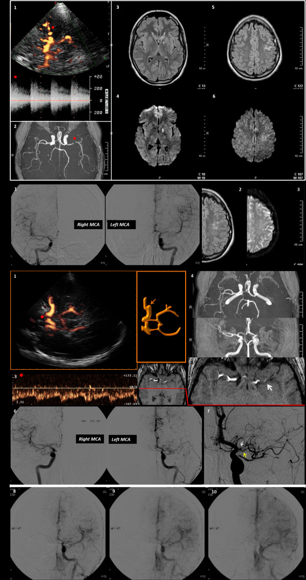
We report the clinical, neurosonological and neuroradiological findings of a young woman, who came to our Stroke Unit because of the abrupt onset of aphasia during her work. An urgent neurosonological examination showed a left M1 MCA stenosis, congruent with the presenting symptoms; magnetic resonance imaging confirmed this finding and identified an acute ischemic lesion on the left MCA territory. The past history of the patient was significant only for a hyperinsulinemic condition, treated with metformine, and a mild overweight. At this time a selective cerebral angiography was not performed because of the patient refusal and she was discharged on antiplatelet and lipid-lowering therapy, having failed to identify autoimmune or inflammatory diseases. Within 1 month, she went back to our attention because of the recurrence of aphasia, lasting about ten minutes. Neuroimaging findings were unchanged, but the patient accepted to undergo a selective cerebral angiography, which showed a mild left distal M1 MCA stenosis.During the follow-up the patient did not experienced any recurrence, but a routine neurosonological examination found an unexpected evolution of the known MCA stenosis, i.e. left M1 MCA occlusion. Neuroradiological imaging did not identify new lesions of the brain parenchyma and a repeated selective cerebral angiography confirmed the left M1 MCA occlusion.
Regardless of the role of metabolic and/or inflammatory factors on the aetiology of the intracranial stenosis in this case, the course of the vessel disease was unexpected and previously unreported in the literature at our knowledge.
颅内大动脉疾病的定位被认为是世界范围内缺血性卒中的主要原因,尽管其全球负担被广泛低估。事实上,它在亚洲人和非裔美国人中更为常见,但在白种人中,颅内狭窄作为缺血性卒中的原因也相对常见。由于颅内狭窄闭塞导致的卒中患者的预后严格取决于再通时间。此外,尽管有一些数据来自于动脉粥样硬化亚型,但与其他病因相比,血管受累的过程在两个方向上(改善或恶化)都是高度动态的。
我们报告了一位年轻女性的临床、神经超声和神经影像学表现,她因工作时突然出现失语症而来到我们的卒中单元。紧急神经超声检查显示左侧 M1 MCA 狭窄,与症状相符;磁共振成像证实了这一发现,并在左侧 MCA 区域识别出一处急性缺血性病变。患者的既往病史仅为高胰岛素血症,用二甲双胍治疗,且有轻度超重。由于患者拒绝,此时未进行选择性脑血管造影,她出院后接受抗血小板和降脂治疗,未能发现自身免疫或炎症性疾病。在 1 个月内,她因失语症再次发作而回到我们的关注中,持续约 10 分钟。神经影像学表现无变化,但患者接受了选择性脑血管造影检查,显示左侧 M1 MCA 轻度远端狭窄。在随访期间,患者未再次发作,但常规神经超声检查发现已知 MCA 狭窄的意外进展,即左侧 M1 MCA 闭塞。神经影像学未发现脑实质的新病变,重复选择性脑血管造影证实左侧 M1 MCA 闭塞。
无论代谢和/或炎症因素在本例颅内狭窄的病因中的作用如何,血管疾病的病程都是意外的,且据我们所知,以前在文献中未曾报道过。