Institut für Neurophysiologie, Medizinische Hochschule Hannover, Hannover, Germany.
PLoS One. 2011;6(12):e29490. doi: 10.1371/journal.pone.0029490. Epub 2011 Dec 19.
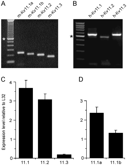
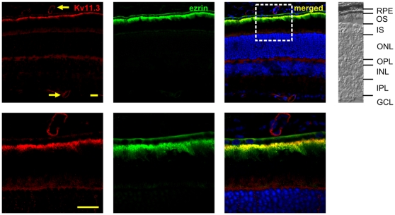
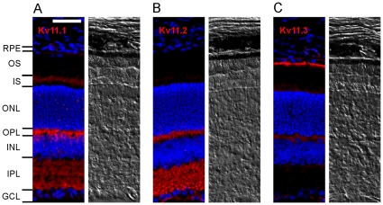
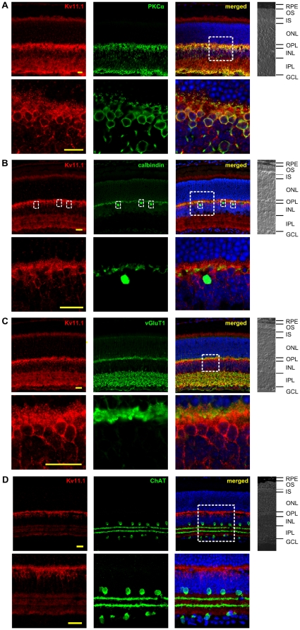
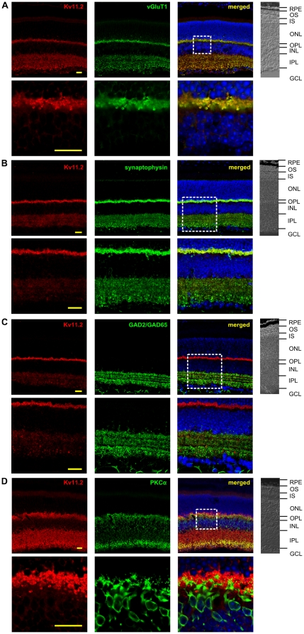
In response to light, most retinal neurons exhibit gradual changes in membrane potential. Therefore K+ channels that mediate threshold currents are well-suited for the fine-tuning of signal transduction. In the present study we demonstrate the expression of the different Kv11 (ether-à-go-go related gene; erg) channel subunits in the human and mouse retina by RT PCR and quantitative PCR, respectively. Immunofluorescence analysis with cryosections of mouse retinae revealed the following local distribution of the three Kv11 subunits: Kv11.1 (m-erg1) displayed the most abundant expression with the strongest immunoreactivity in rod bipolar cells. In addition, immunoreactivity was found in the inner part of the outer plexiform layer (OPL), in the inner plexiform layer (IPL) and in the inner segments of photoreceptors. Immunoreactivity for Kv11.2 (m-erg2) was observed in the outer part of the OPL and throughout the IPL. Double-labeling for vGluT1 or synaptophysin indicated a mainly presynaptic localization of Kv11.2. While no significant staining for Kv11.3 (m-erg3) was detected in the neuronal retina, strong Kv11.3 immunoreactivity was present in the apical membrane of the retinal pigment epithelium. The different expression levels were confirmed by real-time PCR showing almost equal levels of Kv11.1 and Kv11.2, while Kv11.3 mRNA expression was significantly lower. The two main splice variants of Kv11.1, isoforms a and b were detected in comparable levels suggesting a possible formation of cGMP/cGK-sensitive Kv11.1 channels in photoreceptors and rod bipolar cells. Taken together, the immunohistological results revealed different expression patterns of the three Kv11 channels in the mouse retina supposing distinct physiological roles.
对光的反应,大多数视网膜神经元表现出膜电位的逐渐变化。因此,介导阈值电流的 K+通道非常适合精细调节信号转导。在本研究中,我们通过 RT-PCR 和定量 PCR 分别证明了不同 Kv11(醚-去甲肾上腺素相关基因;erg)通道亚基在人眼和鼠眼中的表达。用冷冻切片进行免疫荧光分析显示了三种 Kv11 亚基在小鼠视网膜中的以下局部分布:Kv11.1(m-erg1)表达最丰富,在视杆双极细胞中的免疫反应最强。此外,在外丛状层(OPL)的内部分、内丛状层(IPL)和光感受器的内节中也发现了免疫反应。Kv11.2(m-erg2)的免疫反应在外 OPL 和整个 IPL 中都有观察到。对 vGluT1 或突触小体蛋白的双重标记表明 Kv11.2 主要位于突触前。虽然在神经元视网膜中没有检测到 Kv11.3(m-erg3)的明显染色,但在视网膜色素上皮的顶膜上存在强烈的 Kv11.3 免疫反应。实时 PCR 证实了不同的表达水平,显示 Kv11.1 和 Kv11.2 的水平几乎相等,而 Kv11.3 mRNA 的表达明显较低。Kv11.1 的两种主要剪接变体,a 和 b 型,以可比的水平检测到,这表明在光感受器和视杆双极细胞中可能形成 cGMP/cGK 敏感的 Kv11.1 通道。综上所述,免疫组织化学结果显示了三种 Kv11 通道在小鼠视网膜中的不同表达模式,假设具有不同的生理作用。