Zhou Hongyan, Kimura Kazuhiro, Orita Tomoko, Nishida Teruo, Sonoda Koh-Hei
Department of Ophthalmology, Yamaguchi University Graduate School of Medicine, Ube City, Yamaguchi, Japan.
Mol Vis. 2011;17:3415-22. Epub 2011 Dec 27.
Corneal fibroblasts contribute to collagen remodeling in the corneal stroma in part by mediating collagen degradation. Given that corneal structure is influenced by sex hormone status, we examined the effects of sex hormones on collagen degradation by corneal fibroblasts.
Rabbit corneal fibroblasts were cultured in three-dimensional collagen gels with or without sex hormones including 17β-estradiol, progesterone, testosterone, and dehydroepiandrosterone (DHEA). Collagen degradation was determined by measurement of hydroxyproline after acid hydrolysis. The expression and activity of matrix metalloproteinases (MMPs) were evaluated by immunoblot analysis and gelatin zymography. The phosphorylation of mitogen-activated protein kinases (MAPKs) and the nuclear factor-kappa B (NF-κB) inhibitor NF kappa B Inhibitor-alpha (IκB-α) in corneal fibroblasts was examined by immunoblot analysis. Cell proliferation and viability were evaluated by measurement of bromodeoxyuridine incorporation and the release of lactate dehydrogenase, respectively.
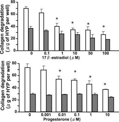
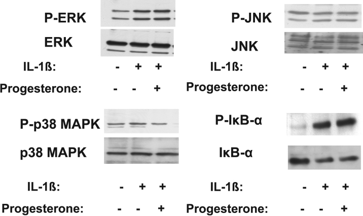
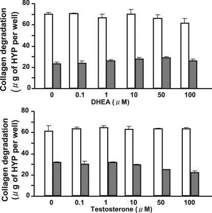
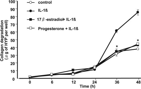
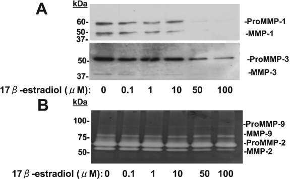
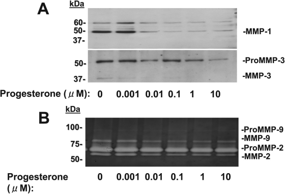
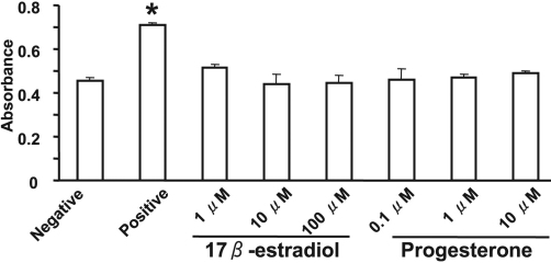
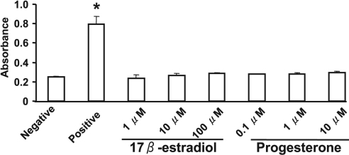
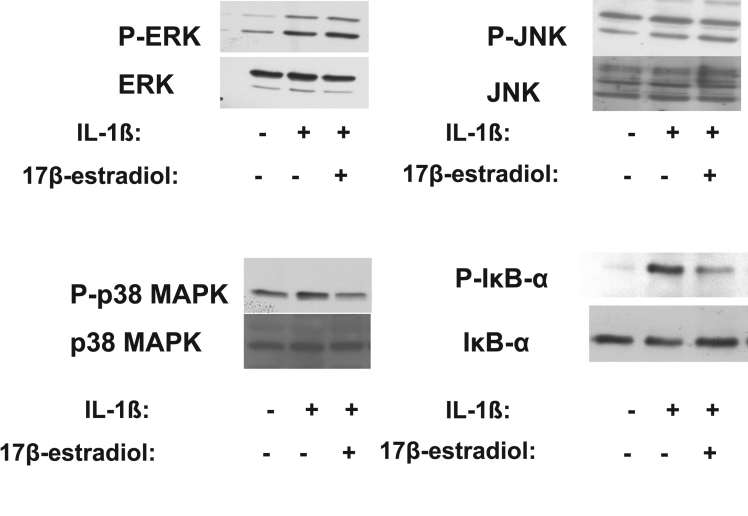
17β-Estradiol and progesterone each inhibited interleukin (IL)-1β-induced collagen degradation by corneal fibroblasts in a concentration-dependent manner, whereas testosterone and DHEA had no such effect. MMP expression and activation in corneal fibroblasts exposed to IL-1β were also inhibited by 17β-estradiol and progesterone. These female sex hormones did not affect cell proliferation or viability. Both 17β-estradiol and progesterone inhibited the IL-1β-induced phosphorylation of p38 MAPK without affecting that of the MAPKs extracellular Signal-regulated Kinase (ERK) or c-jun N-terminal kinase (JNK). 17β-Estradiol also inhibited the IL-1β-induced phosphorylation of IκB-α.
17β-Estradiol and progesterone inhibited MMP expression and activity in IL-1β-stimulated corneal fibroblasts and thereby suppressed collagen degradation by these cells.
角膜成纤维细胞部分通过介导胶原蛋白降解,对角膜基质中的胶原蛋白重塑起作用。鉴于角膜结构受性激素状态影响,我们研究了性激素对角膜成纤维细胞胶原蛋白降解的作用。
兔角膜成纤维细胞在含或不含性激素(包括17β-雌二醇、孕酮、睾酮和脱氢表雄酮(DHEA))的三维胶原凝胶中培养。通过酸水解后测量羟脯氨酸来确定胶原蛋白降解情况。通过免疫印迹分析和明胶酶谱法评估基质金属蛋白酶(MMPs)的表达和活性。通过免疫印迹分析检测角膜成纤维细胞中丝裂原活化蛋白激酶(MAPKs)的磷酸化以及核因子-κB(NF-κB)抑制剂NFκB抑制剂α(IκB-α)。分别通过测量溴脱氧尿苷掺入量和乳酸脱氢酶释放量来评估细胞增殖和活力。
17β-雌二醇和孕酮均以浓度依赖性方式抑制白细胞介素(IL)-1β诱导的角膜成纤维细胞胶原蛋白降解,而睾酮和DHEA则无此作用。暴露于IL-1β的角膜成纤维细胞中MMP的表达和活化也受到17β-雌二醇和孕酮的抑制。这些雌性激素不影响细胞增殖或活力。17β-雌二醇和孕酮均抑制IL-1β诱导的p38 MAPK磷酸化,而不影响细胞外信号调节激酶(ERK)或c-jun氨基末端激酶(JNK)的磷酸化。17β-雌二醇还抑制IL-1β诱导的IκB-α磷酸化。
17β-雌二醇和孕酮抑制IL-1β刺激的角膜成纤维细胞中MMP的表达和活性,从而抑制这些细胞的胶原蛋白降解。