Lin Haotian, Chen Weirong, Luo Lixia, Wu Changrui, Wang Qilin, Liu Yizhi
State Key Laboratory of Ophthalmology, Zhongshan Ophthalmic Center, Sun Yat-sen University, Guangzhou, China.
Mol Vis. 2011;17:3450-7. Epub 2011 Dec 28.
This research was conducted to make a primary culture of human retinal capillary endothelial cells (HRCEC) and to study the cytotoxic effect of human immunodeficiency virus-1 (envelope) glycoprotein 120 (HIV-1 gp120) on cultured HRCEC.
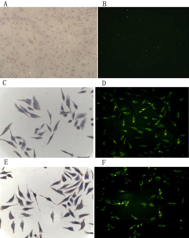
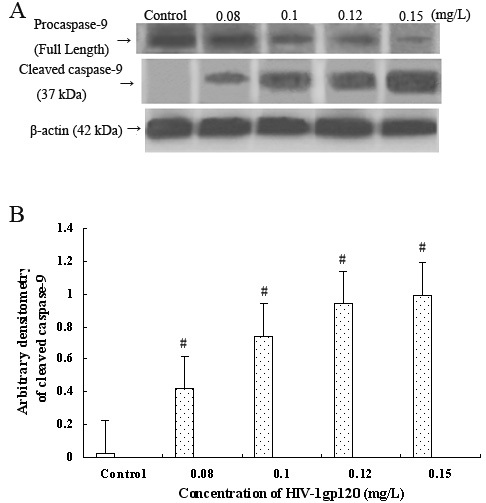
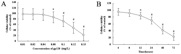
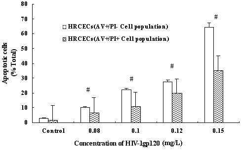
HRCEC were isolated and primarily cultured as dissociated single cell cultures. Immunohistochemistry and immunofluorescence were used to identify specific markers of HRCEC and to reveal HIV-1 gp120 related receptors (cluster of differentiation 4 [CD4], C-X-C chemokine receptor type 4 [CXCR4], and C-C chemokine receptor type 5 [CCR5]). The 3-[4,5-dimethylthiazol-2-yl]-2,5-diphenyl tetrazolium bromide (MTT) assay was used to demonstrate the effect of HIV-1 gp120 on cell viability at seven different concentrations (0.01-0.15 mg/l) for 24 h or at a fixed concentration of 0.08 mg/l for varying time intervals (4-72 h). After 0.08, 0.1, 0.12, and 0.15 mg/l HIV-1 gp120 were applied to HRCEC for 24 h, cell apoptotic rates and the mitochondrial membrane potential were measured with flow cytometry; pro-caspase-9 and cleaved caspase-9 were evaluated with immunoblotting. Under each research condition, 0.15 mg/l of HIV-1 gp120 mutated proteins (423 I/P) were used as controls.
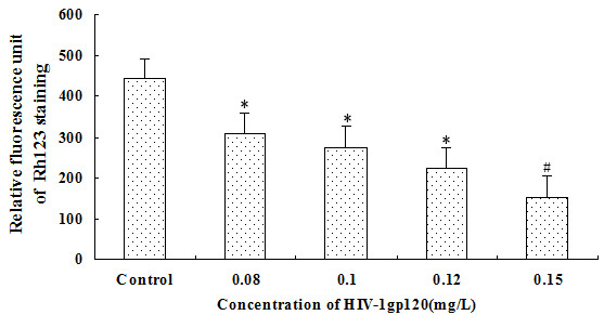
Primary cultures of pure HRCEC were established, and the cells were characterized with their specific markers. HIV-1 gp120 receptors CXCR4 and CCR5 were found on the cell surface of HRCEC; however, CD4 was negative. Treatment of HRCEC with HIV-1 gp120 at concentrations <0.08 mg/l did not influence cell viability. However, a concentration- and time-dependent increase of HIV-1 gp120-induced cell inhibition was demonstrated with MTT, when the concentration of HIV-1 gp120 was more than 0.08 mg/l (r=-0.763, p<0.01). With increasing concentrations of HIV-1 gp120, the numbers of apoptotic cells and expression of cleaved caspase-9 protein increased, but Rho123 staining mitochondrial membrane potential decreased.
HIV-1 gp120 assistant receptors CXCR4 and CCR5 are expressed on the cell surface of HRCEC, and HIV-1 gp120 can inhibit cell viability and induce apoptosis of HRCEC. The mitochondrial pathway is probably involved in HIV-1 gp120-induced apoptosis of HRCEC, but the specific mechanisms remain to be uncovered.
本研究旨在进行人视网膜毛细血管内皮细胞(HRCEC)的原代培养,并研究人类免疫缺陷病毒1型(包膜)糖蛋白120(HIV-1 gp120)对培养的HRCEC的细胞毒性作用。
将HRCEC分离并作为解离的单细胞培养物进行原代培养。采用免疫组织化学和免疫荧光法鉴定HRCEC的特异性标志物,并揭示HIV-1 gp120相关受体(分化簇4 [CD4]、C-X-C趋化因子受体4型[CXCR4]和C-C趋化因子受体5型[CCR5])。采用3-[4,5-二甲基噻唑-2-基]-2,5-二苯基溴化四氮唑(MTT)法,以7种不同浓度(0.01 - 0.15 mg/l)处理24小时或固定浓度0.08 mg/l处理不同时间间隔(4 - 72小时),以证明HIV-1 gp120对细胞活力的影响。在将0.08、0.1、0.12和0.15 mg/l的HIV-1 gp120应用于HRCEC 24小时后,用流式细胞术测量细胞凋亡率和线粒体膜电位;用免疫印迹法评估前半胱天冬酶-9和裂解的半胱天冬酶-9。在每种研究条件下,使用0.15 mg/l的HIV-1 gp120突变蛋白(423 I/P)作为对照。
建立了纯HRCEC的原代培养物,并用其特异性标志物对细胞进行了表征。在HRCEC的细胞表面发现了HIV-1 gp120受体CXCR4和CCR5;然而,CD4为阴性。用浓度<0.08 mg/l的HIV-1 gp120处理HRCEC不影响细胞活力。然而,MTT法显示,当HIV-1 gp120浓度超过0.08 mg/l时,HIV-1 gp120诱导的细胞抑制呈浓度和时间依赖性增加(r = -0.763,p<0.01)。随着HIV-1 gp120浓度的增加,凋亡细胞数量和裂解的半胱天冬酶-9蛋白表达增加,但罗丹明123染色的线粒体膜电位降低。
HIV-1 gp120辅助受体CXCR4和CCR5在HRCEC的细胞表面表达,HIV-1 gp120可抑制HRCEC的细胞活力并诱导其凋亡。线粒体途径可能参与HIV-1 gp120诱导的HRCEC凋亡,但具体机制仍有待揭示。