• 文献检索
  • 文档翻译
  • 深度研究
  • 学术资讯
  • Suppr Zotero 插件Zotero 插件
  • 邀请有礼
  • 套餐&价格
  • 历史记录
应用&插件
Suppr Zotero 插件Zotero 插件浏览器插件Mac 客户端Windows 客户端微信小程序
定价
高级版会员购买积分包购买API积分包
服务
文献检索文档翻译深度研究API 文档MCP 服务
关于我们
关于 Suppr公司介绍联系我们用户协议隐私条款
关注我们

Suppr 超能文献

核心技术专利:CN118964589B侵权必究
粤ICP备2023148730 号-1Suppr @ 2026

文献检索

告别复杂PubMed语法,用中文像聊天一样搜索,搜遍4000万医学文献。AI智能推荐,让科研检索更轻松。

立即免费搜索

文件翻译

保留排版,准确专业,支持PDF/Word/PPT等文件格式,支持 12+语言互译。

免费翻译文档

深度研究

AI帮你快速写综述,25分钟生成高质量综述,智能提取关键信息,辅助科研写作。

立即免费体验

铁螯合降低大鼠脑出血后脑内铁水平但不改善预后。

Ferric iron chelation lowers brain iron levels after intracerebral hemorrhage in rats but does not improve outcome.

机构信息

Dept. of Neurosurgery, Stanford University School of Medicine, USA.

出版信息

Exp Neurol. 2012 Mar;234(1):136-43. doi: 10.1016/j.expneurol.2011.12.030. Epub 2011 Dec 27.

DOI:10.1016/j.expneurol.2011.12.030
PMID:22226595
原文链接:https://pmc.ncbi.nlm.nih.gov/articles/PMC3848975/
Abstract

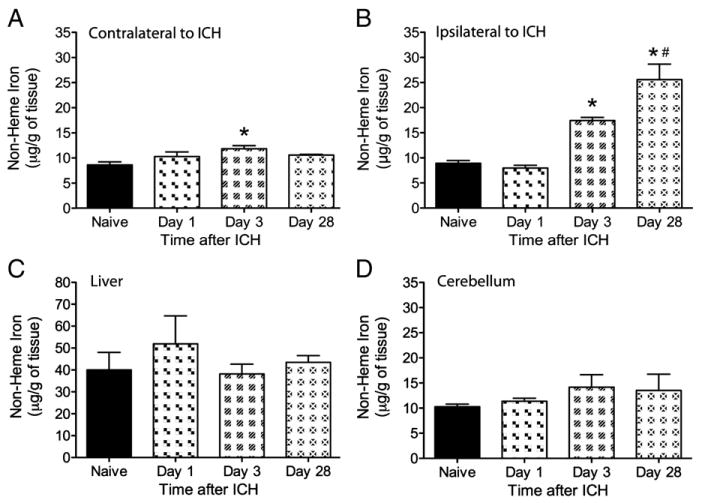
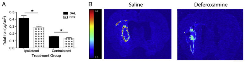
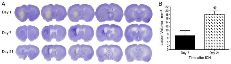
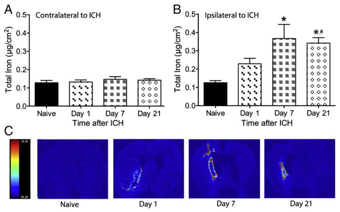
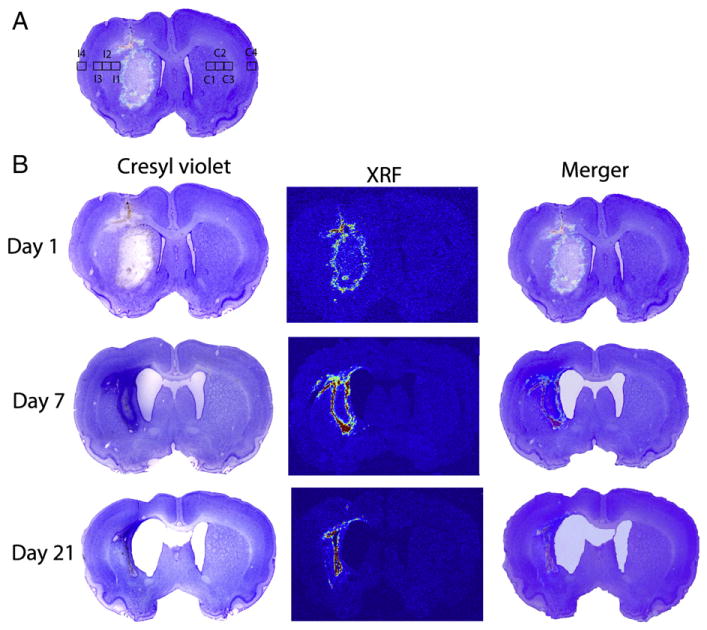
Iron-mediated free radical damage contributes to secondary damage after intracerebral hemorrhage (ICH). Iron is released from heme after hemoglobin breakdown and accumulates in the parenchyma over days and then persists in the brain for months (e.g., hemosiderin). This non-heme iron has been linked to cerebral edema and cell death. Deferoxamine, a ferric iron chelator, has been shown to mitigate iron-mediated damage, but results vary with less protection in the collagenase model of ICH. This study used rapid-scanning X-ray fluorescence (RS-XRF), a synchrotron-based imaging technique, to spatially map total iron and other elements (zinc, calcium and sulfur) at three survival times after collagenase-induced ICH in rats. Total iron was compared to levels of non-heme iron determined by a Ferrozine-based spectrophotometry assay in separate animals. Finally, using RS-XRF we measured iron levels in ICH rats treated with deferoxamine versus saline. The non-heme iron assay showed elevations in injured striatum at 3 days and 4 weeks post-ICH, but not at 1 day. RS-XRF also detected significantly increased iron levels at comparable times, especially notable in the peri-hematoma zone. Changes in other elements were observed in some animals, but these were inconsistent among animals. Deferoxamine diminished total parenchymal iron levels but did not attenuate neurological deficits or lesion volume at 7 days. In summary, ICH significantly increased non-heme and total iron levels. We evaluated the latter and found it to be significantly lowered by deferoxamine, but its failure to attenuate injury or functional impairment in this model raises concern about successful translation to patients.

摘要

铁介导的自由基损伤导致脑出血(ICH)后的继发性损伤。血红蛋白分解后,铁从血红素中释放出来,并在数天内在实质中积累,然后在大脑中持续数月(例如含铁血黄素)。这种非血红素铁与脑水肿和细胞死亡有关。去铁胺是一种三价铁螯合剂,已被证明可以减轻铁介导的损伤,但在胶原酶诱导的 ICH 模型中,其保护作用较差。本研究使用快速扫描 X 射线荧光(RS-XRF),一种基于同步加速器的成像技术,在胶原酶诱导的 ICH 后大鼠三个存活时间点空间映射总铁和其他元素(锌、钙和硫)。总铁与通过 Ferrozine 分光光度法测定的非血红素铁水平进行比较。最后,使用 RS-XRF 我们测量了用去铁胺与生理盐水治疗的 ICH 大鼠的铁水平。非血红素铁测定法显示,ICH 后 3 天和 4 周时损伤纹状体中的非血红素铁水平升高,但 1 天时没有升高。RS-XRF 还在可比时间检测到明显增加的铁水平,尤其是在血肿周围区域。在一些动物中观察到其他元素的变化,但这些变化在动物之间不一致。去铁胺降低了总实质铁水平,但在 7 天时并未减轻神经功能缺损或病变体积。总之,ICH 显著增加了非血红素和总铁水平。我们评估了后者,发现去铁胺显著降低了后者,但它未能减轻该模型中的损伤或功能障碍,这引起了对成功转化为患者的关注。

https://cdn.ncbi.nlm.nih.gov/pmc/blobs/400e/3848975/040fadbfecfd/nihms3545f7.jpg
https://cdn.ncbi.nlm.nih.gov/pmc/blobs/400e/3848975/5e9a9fa10a88/nihms3545f1.jpg
https://cdn.ncbi.nlm.nih.gov/pmc/blobs/400e/3848975/65c2718864c0/nihms3545f2.jpg
https://cdn.ncbi.nlm.nih.gov/pmc/blobs/400e/3848975/422996de0cd4/nihms3545f3.jpg
https://cdn.ncbi.nlm.nih.gov/pmc/blobs/400e/3848975/d2d7e86915fc/nihms3545f4.jpg
https://cdn.ncbi.nlm.nih.gov/pmc/blobs/400e/3848975/f0048d2a3943/nihms3545f5.jpg
https://cdn.ncbi.nlm.nih.gov/pmc/blobs/400e/3848975/51e54f0771b7/nihms3545f6.jpg
https://cdn.ncbi.nlm.nih.gov/pmc/blobs/400e/3848975/040fadbfecfd/nihms3545f7.jpg

相似文献

1
Ferric iron chelation lowers brain iron levels after intracerebral hemorrhage in rats but does not improve outcome.铁螯合降低大鼠脑出血后脑内铁水平但不改善预后。
Exp Neurol. 2012 Mar;234(1):136-43. doi: 10.1016/j.expneurol.2011.12.030. Epub 2011 Dec 27.
2
Failure of deferoxamine, an iron chelator, to improve outcome after collagenase-induced intracerebral hemorrhage in rats.铁螯合剂去铁胺治疗胶原酶诱导的大鼠脑出血失败。
Brain Res. 2010 Jan 14;1309:95-103. doi: 10.1016/j.brainres.2009.10.058. Epub 2009 Oct 30.
3
Activation of c-Jun-N-terminal kinase in a rat model of intracerebral hemorrhage: the role of iron.脑出血大鼠模型中c-Jun氨基末端激酶的激活:铁的作用
Neurosci Res. 2009 Feb;63(2):100-5. doi: 10.1016/j.neures.2008.10.013. Epub 2008 Nov 30.
4
Deferoxamine therapy for intracerebral hemorrhage.去铁胺治疗脑出血。
Acta Neurochir Suppl. 2008;105:3-6. doi: 10.1007/978-3-211-09469-3_1.
5
Deferoxamine reduces neuronal death and hematoma lysis after intracerebral hemorrhage in aged rats.去铁胺可减少老年大鼠脑出血后的神经元死亡和血肿溶解。
Transl Stroke Res. 2013 Oct;4(5):546-53. doi: 10.1007/s12975-013-0270-5.
6
The effects of deferoxamine on inhibition for microglia activation and protection of secondary nerve injury after intracerebral hemorrhage in rats.去铁胺对大鼠脑出血后小胶质细胞激活的抑制作用及继发性神经损伤的保护作用
Pak J Pharm Sci. 2016 May;29(3 Suppl):1087-93.
7
Deferoxamine-induced attenuation of brain edema and neurological deficits in a rat model of intracerebral hemorrhage.去铁胺减轻大鼠脑出血模型中的脑水肿和神经功能缺损
Neurosurg Focus. 2003 Oct 15;15(4):ECP4. doi: 10.3171/foc.2003.15.4.10.
8
Iron accumulation and DNA damage in a pig model of intracerebral hemorrhage.脑出血猪模型中的铁蓄积与DNA损伤
Acta Neurochir Suppl. 2011;111:123-8. doi: 10.1007/978-3-7091-0693-8_20.
9
Brain edema after intracerebral hemorrhage in rats: the role of iron overload and aquaporin 4.大鼠脑出血后的脑水肿:铁过载和水通道蛋白4的作用
J Neurosurg. 2009 Mar;110(3):462-8. doi: 10.3171/2008.4.JNS17512.
10
Deferoxamine-induced attenuation of brain edema and neurological deficits in a rat model of intracerebral hemorrhage.去铁胺减轻大鼠脑出血模型中的脑水肿和神经功能缺损
J Neurosurg. 2004 Apr;100(4):672-8. doi: 10.3171/jns.2004.100.4.0672.

引用本文的文献

1
Characteristics of Scar Formation After Intracerebral Hemorrhage in Aged Rats: Effects of Deferoxamine.老年大鼠脑出血后瘢痕形成的特征:去铁胺的作用
Cells. 2025 Jul 22;14(15):1127. doi: 10.3390/cells14151127.
2
Iron Neurotoxicity and Protection by Deferoxamine in Intracerebral Hemorrhage.脑出血中铁的神经毒性及去铁胺的保护作用
Front Mol Neurosci. 2022 Jun 16;15:927334. doi: 10.3389/fnmol.2022.927334. eCollection 2022.
3
Minimally Invasive Surgery for ICH Evacuation Combined With Deferoxamine Treatment Increased Perihematomal Claudin-5 and ZO-1 Expression Levels and Decreased BBB Permeability in Rabbits.

本文引用的文献

1
A critical appraisal of experimental intracerebral hemorrhage research.对实验性脑出血研究的批判性评价。
J Cereb Blood Flow Metab. 2012 Apr;32(4):612-27. doi: 10.1038/jcbfm.2012.8. Epub 2012 Feb 1.
2
Efficacy of the lipid-soluble iron chelator 2,2'-dipyridyl against hemorrhagic brain injury.脂溶性铁螯合剂 2,2'-联吡啶对脑出血损伤的疗效。
Neurobiol Dis. 2012 Jan;45(1):388-94. doi: 10.1016/j.nbd.2011.08.028. Epub 2011 Sep 10.
3
Safety and tolerability of deferoxamine mesylate in patients with acute intracerebral hemorrhage.
微创脑出血清除术联合去铁胺治疗可提高兔血肿周围紧密连接蛋白5和闭锁小带蛋白1的表达水平并降低血脑屏障通透性。
Front Neurol. 2022 Mar 3;13:835494. doi: 10.3389/fneur.2022.835494. eCollection 2022.
4
Mechanism of Ferroptosis and Its Relationships With Other Types of Programmed Cell Death: Insights for Potential Interventions After Intracerebral Hemorrhage.铁死亡的机制及其与其他类型程序性细胞死亡的关系:对脑出血后潜在干预措施的见解
Front Neurosci. 2020 Nov 13;14:589042. doi: 10.3389/fnins.2020.589042. eCollection 2020.
5
PEGylation of Deferoxamine for Improving the Stability, Cytotoxicity, and Iron-Overload in an Experimental Stroke Model in Rats.去铁胺的聚乙二醇化修饰对改善大鼠实验性脑卒中模型中的稳定性、细胞毒性和铁过载的作用
Front Bioeng Biotechnol. 2020 Sep 25;8:592294. doi: 10.3389/fbioe.2020.592294. eCollection 2020.
6
Tracking elemental changes in an ischemic stroke model with X-ray fluorescence imaging.用 X 射线荧光成像技术追踪缺血性中风模型中的元素变化。
Sci Rep. 2020 Oct 20;10(1):17868. doi: 10.1038/s41598-020-74698-2.
7
Perihematomal brain tissue iron concentration measurement by MRI in patients with intracerebral hemorrhage.磁共振成像测量脑出血患者血肿周围脑组织铁浓度。
CNS Neurosci Ther. 2020 Sep;26(9):896-901. doi: 10.1111/cns.13395. Epub 2020 May 21.
8
Pathophysiological Mechanisms and Potential Therapeutic Targets in Intracerebral Hemorrhage.脑出血的病理生理机制及潜在治疗靶点
Front Pharmacol. 2019 Sep 19;10:1079. doi: 10.3389/fphar.2019.01079. eCollection 2019.
9
The Involvement of Iron in Traumatic Brain Injury and Neurodegenerative Disease.铁在创伤性脑损伤和神经退行性疾病中的作用
Front Neurosci. 2018 Dec 20;12:981. doi: 10.3389/fnins.2018.00981. eCollection 2018.
10
Prolonged Blood-Brain Barrier Injury Occurs After Experimental Intracerebral Hemorrhage and Is Not Acutely Associated with Additional Bleeding.实验性脑出血后会发生持续性血脑屏障损伤,且与额外出血并无急性相关性。
Transl Stroke Res. 2019 Jun;10(3):287-297. doi: 10.1007/s12975-018-0636-9. Epub 2018 Jun 14.
甲磺酸去铁胺治疗急性脑出血患者的安全性和耐受性。
Stroke. 2011 Nov;42(11):3067-74. doi: 10.1161/STROKEAHA.111.617589. Epub 2011 Aug 25.
4
Experimental intracerebral hemorrhage: avoiding pitfalls in translational research.实验性脑出血:避免转化研究中的陷阱。
J Cereb Blood Flow Metab. 2011 Nov;31(11):2135-51. doi: 10.1038/jcbfm.2011.124. Epub 2011 Aug 24.
5
Zinc: new clues to diverse roles in brain ischemia.锌:脑缺血中多种作用的新线索。
Trends Pharmacol Sci. 2011 Aug;32(8):480-6. doi: 10.1016/j.tips.2011.04.001. Epub 2011 May 27.
6
Molecular pathophysiology of cerebral hemorrhage: secondary brain injury.脑出血的分子病理生理学:继发性脑损伤。
Stroke. 2011 Jun;42(6):1781-6. doi: 10.1161/STROKEAHA.110.596718. Epub 2011 Apr 28.
7
Treatment of intracerebral hemorrhage in animal models: meta-analysis.脑出血动物模型的治疗:荟萃分析。
Ann Neurol. 2011 Feb;69(2):389-99. doi: 10.1002/ana.22243.
8
Iron toxicity in mice with collagenase-induced intracerebral hemorrhage.胶原酶诱导脑出血小鼠的铁毒性。
J Cereb Blood Flow Metab. 2011 May;31(5):1243-50. doi: 10.1038/jcbfm.2010.209. Epub 2010 Nov 24.
9
Towards a unifying, systems biology understanding of large-scale cellular death and destruction caused by poorly liganded iron: Parkinson's, Huntington's, Alzheimer's, prions, bactericides, chemical toxicology and others as examples.从系统生物学角度理解配体结合不良的铁导致的大规模细胞死亡和破坏:以帕金森病、亨廷顿病、阿尔茨海默病、朊病毒、杀菌剂、化学毒理学等为例。
Arch Toxicol. 2010 Nov;84(11):825-89. doi: 10.1007/s00204-010-0577-x. Epub 2010 Aug 17.
10
T2* magnetic resonance imaging sequences reflect brain tissue iron deposition following intracerebral hemorrhage.T2* 磁共振成像序列反映脑出血后脑组织铁沉积。
Transl Stroke Res. 2010 Mar 1;1(1):31-4. doi: 10.1007/s12975-009-0008-6.