The Children’s Hospital at Westmead, Westmead, Australia.
Diabetes Care. 2012 Mar;35(3):599-604. doi: 10.2337/dc11-1177. Epub 2012 Jan 16.
To examine the relationship between retinal vascular geometry parameters and development of incident renal dysfunction in young people with type 1 diabetes.
This was a prospective cohort study of 511 adolescents with type 1 diabetes of at least 2 years duration, with normal albumin excretion rate (AER) and no retinopathy at baseline while attending an Australian tertiary-care hospital. AER was quantified using three overnight, timed urine specimen collections and early renal dysfunction was defined as AER >7.5 μg/min. Retinal vascular geometry (including length-to-diameter ratio [LDR] and simple tortuosity [ST]) was quantified from baseline retinal photographs. Generalized estimating equations were used to examine the relationship between incident renal dysfunction and baseline venular LDR and ST, adjusting for age, diabetes duration, glycated hemoglobin (A1C), blood pressure (BP), BMI, and cholesterol.
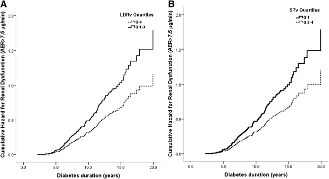
Diabetes duration at baseline was 4.8 (IQR 3.3-7.5) years. After a median 3.7 (2.3-5.7) years follow-up, 34% of participants developed incident renal dysfunction. In multivariate analysis, higher retinal venular LDR (odds ratio 1.7, 95% CI 1.2-2.4; quartile 4 vs. 1-3) and lower venular ST (1.6, 1.1-2.2; quartile 1 vs. 2-4) predicted incident renal dysfunction.
Retinal venular geometry independently predicted incident renal dysfunction in young people with type 1 diabetes. These noninvasive retinal measures may help to elucidate early mechanistic pathways for microvascular complications. Retinal venular geometry may be a useful tool to identify individuals at high risk of renal disease early in the course of diabetes.
研究视网膜血管几何参数与 1 型糖尿病青少年新发肾功能障碍的关系。
这是一项对 511 名至少患有 2 年 1 型糖尿病、基线时正常白蛋白排泄率(AER)且无视网膜病变的青少年进行的前瞻性队列研究,这些患者在澳大利亚三级护理医院就诊。通过三次过夜、定时尿液标本采集来定量 AER,早期肾功能障碍定义为 AER>7.5μg/min。从基线视网膜照片中量化视网膜血管几何形状(包括长度与直径比[LDR]和简单迂曲[ST])。使用广义估计方程来检验新发肾功能障碍与基线静脉 LDR 和 ST 之间的关系,调整年龄、糖尿病病程、糖化血红蛋白(A1C)、血压(BP)、BMI 和胆固醇。
基线时的糖尿病病程为 4.8(IQR 3.3-7.5)年。在中位数为 3.7(2.3-5.7)年的随访后,34%的参与者出现新发肾功能障碍。在多变量分析中,较高的视网膜静脉 LDR(比值比 1.7,95%CI 1.2-2.4;四分位 4 与 1-3)和较低的静脉 ST(1.6,1.1-2.2;四分位 1 与 2-4)预测了新发肾功能障碍。
视网膜静脉几何形状独立预测了 1 型糖尿病青少年新发肾功能障碍。这些非侵入性的视网膜测量方法可能有助于阐明微血管并发症的早期机制途径。视网膜静脉几何形状可能是一种有用的工具,可在糖尿病病程早期识别出患有肾病高风险的个体。