Murakami Yuichi, Okazaki Yoshimasa, Okayama Shinji, Fujihira Shiro, Noto Takahisa, Nakatsuji Shunji, Oishi Yuji
Toxicologic Pathology, Drug Safety Research Labs., Astellas Pharma Inc., 2-1-6 Kashima, Yodogawa-ku, Osaka 532-8514, Japan.
J Toxicol Pathol. 2010 Jun;23(2):85-9. doi: 10.1293/tox.23.85. Epub 2010 Jun 30.
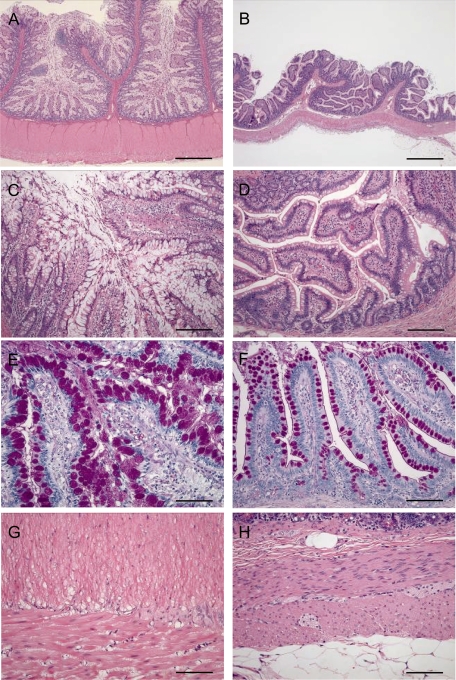
We report here the interesting case of a 5-year-old male cynomolgus monkey with goblet cell hyperplasia and thickening of the muscular layer throughout the small intestine without exhibiting any clinical symptoms. Necropsy examination showed diffuse thickening of the intestinal wall from the jejunum to the ileum, with an appearance likened to a rubber tube. Histopathologically, marked thickening was observed in both the mucosal and muscular layers in the jejunum and ileum, and slight thickening was observed in the duodenum. Goblet cell hyperplasia with extension of the circular folds and villi was prominently observed. The mucosal surface was covered with a thick mucus layer containing desquamated mucosal epithelial cells, and both the inner and outer muscular layers were markedly thickened due to smooth muscle hypertrophy. Neither macroscopic nor histopathological examination identified any causative factors, such as infection, enteritis and intestinal stenosis, or obstruction that may have caused development of this lesion. Given these observations, this case may simply be considered of spontaneous goblet cell hyperplasia and muscular layer thickening in the small intestine of a cynomolgus monkey.
我们在此报告一例有趣的病例,一只5岁雄性食蟹猴,其小肠出现杯状细胞增生和肌层增厚,但未表现出任何临床症状。尸检显示,从空肠到回肠肠壁弥漫性增厚,外观类似橡胶管。组织病理学检查发现,空肠和回肠的黏膜层和肌层均有明显增厚,十二指肠有轻微增厚。显著观察到杯状细胞增生,并伴有环形皱襞和绒毛延长。黏膜表面覆盖着一层厚厚的黏液层,其中含有脱落的黏膜上皮细胞,由于平滑肌肥大,内、外肌层均明显增厚。宏观和组织病理学检查均未发现任何可能导致该病变发展的致病因素,如感染、肠炎、肠道狭窄或梗阻。基于这些观察结果,该病例可能仅被视为食蟹猴小肠自发性杯状细胞增生和肌层增厚。