• 文献检索
  • 文档翻译
  • 深度研究
  • 学术资讯
  • Suppr Zotero 插件Zotero 插件
  • 邀请有礼
  • 套餐&价格
  • 历史记录
应用&插件
Suppr Zotero 插件Zotero 插件浏览器插件Mac 客户端Windows 客户端微信小程序
定价
高级版会员购买积分包购买API积分包
服务
文献检索文档翻译深度研究API 文档MCP 服务
关于我们
关于 Suppr公司介绍联系我们用户协议隐私条款
关注我们

Suppr 超能文献

核心技术专利:CN118964589B侵权必究
粤ICP备2023148730 号-1Suppr @ 2026

文献检索

告别复杂PubMed语法,用中文像聊天一样搜索,搜遍4000万医学文献。AI智能推荐,让科研检索更轻松。

立即免费搜索

文件翻译

保留排版,准确专业,支持PDF/Word/PPT等文件格式,支持 12+语言互译。

免费翻译文档

深度研究

AI帮你快速写综述,25分钟生成高质量综述,智能提取关键信息,辅助科研写作。

立即免费体验

印度人群下颌第一恒磨牙的根管形态

Root canal morphology of mandibular first permanent molars in an Indian population.

作者信息

Chourasia Hemant Ramesh, Meshram Ganesh K, Warhadpande Manjusha, Dakshindas Darshan

机构信息

Department of Conservative Dentistry and Endodontics, Peoples College of Dental Sciences and Research Centre, Bhanpur, Bhopal 462037, India.

出版信息

Int J Dent. 2012;2012:745152. doi: 10.1155/2012/745152. Epub 2012 Jan 11.

DOI:10.1155/2012/745152
PMID:22287965
原文链接:https://pmc.ncbi.nlm.nih.gov/articles/PMC3263621/
Abstract

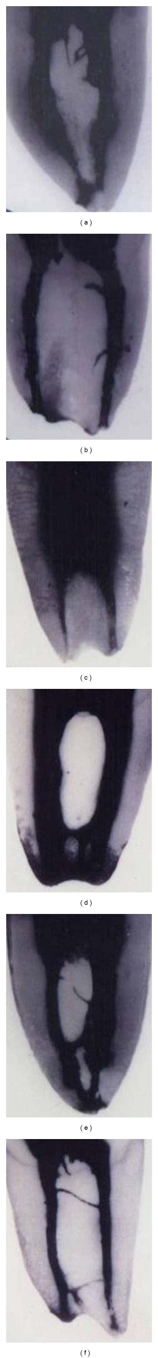
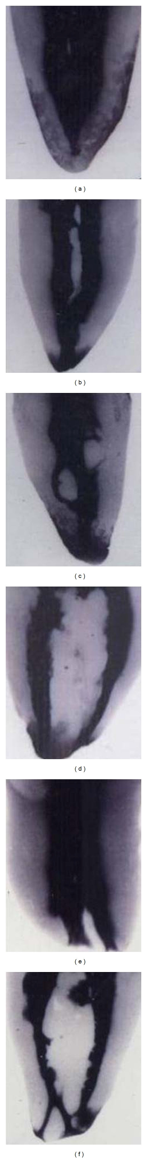
An in vitro study was performed to determine the number of roots, root canals per tooth, root canal configurations, and frequency of isthmi and apical deltas in mandibular first permanent molars in an Indian population. Hundred and fifty mandibular first permanent molars were collected and subjected to clearing technique. The cleared teeth were examined in a stereomicroscope under 7.5x magnifications. The canal configurations were categorized using Vertucci's classification. Overall 94.6% of the mandibular first molars had two roots, and 5.3% had extradistal roots (distolingual root). In addition, 64% of the specimens had three root canals, and 36% had four root canals. The most common canal configurations of mesial and distal roots were Vertucci type IV (54%) and type I (65.3%), respectively. Clinician should be aware of the complex root canal morphology of mandibular first molars among the Indian population before and during the root canal treatment.

摘要

进行了一项体外研究,以确定印度人群下颌第一恒磨牙的牙根数量、每颗牙齿的根管数量、根管形态以及峡部和根尖分歧的发生率。收集了150颗下颌第一恒磨牙并采用透明技术进行处理。在体视显微镜下以7.5倍放大倍数检查透明后的牙齿。根管形态采用韦尔图奇分类法进行分类。总体而言,94.6%的下颌第一磨牙有两个牙根,5.3%有额外远中根(远舌根)。此外,64%的标本有三个根管,36%有四个根管。近中根和远中根最常见的根管形态分别为韦尔图奇IV型(54%)和I型(65.3%)。临床医生在根管治疗前和治疗过程中应了解印度人群下颌第一磨牙复杂的根管形态。

https://cdn.ncbi.nlm.nih.gov/pmc/blobs/a914/3263621/47dcc1ffb216/IJD2012-745152.002.jpg
https://cdn.ncbi.nlm.nih.gov/pmc/blobs/a914/3263621/e321b6ddf6cf/IJD2012-745152.001.jpg
https://cdn.ncbi.nlm.nih.gov/pmc/blobs/a914/3263621/47dcc1ffb216/IJD2012-745152.002.jpg
https://cdn.ncbi.nlm.nih.gov/pmc/blobs/a914/3263621/e321b6ddf6cf/IJD2012-745152.001.jpg
https://cdn.ncbi.nlm.nih.gov/pmc/blobs/a914/3263621/47dcc1ffb216/IJD2012-745152.002.jpg

相似文献

1
Root canal morphology of mandibular first permanent molars in an Indian population.印度人群下颌第一恒磨牙的根管形态
Int J Dent. 2012;2012:745152. doi: 10.1155/2012/745152. Epub 2012 Jan 11.
2
Root and canal morphology of mandibular second molars in an Indian population.下颌第二磨牙的根管和根形态在印度人群中的研究。
J Endod. 2010 Aug;36(8):1319-22. doi: 10.1016/j.joen.2010.04.001. Epub 2010 May 23.
3
Root canal morphology of South Asian Indian mandibular first, second, and third molar: A dye penetration and clearing study.南亚印度人下颌第一、第二和第三磨牙的根管形态:一项染料渗透与透明研究。
J Conserv Dent. 2020 May-Jun;23(3):284-288. doi: 10.4103/JCD.JCD_379_20. Epub 2020 Dec 4.
4
Investigation of the root canal configuration of mandibular first molars in a Taiwan Chinese population.台湾华裔人群下颌第一磨牙根管形态的研究。
Int Endod J. 2009 Nov;42(11):1044-9. doi: 10.1111/j.1365-2591.2009.01619.x.
5
Anatomical variations and bilateral symmetry of roots and root canal system of mandibular first permanent molars in Saudi Arabian population utilizing cone- beam computed tomography.利用锥形束计算机断层扫描技术研究沙特阿拉伯人群下颌第一恒磨牙牙根及根管系统的解剖变异和双侧对称性
Saudi Dent J. 2019 Oct;31(4):481-486. doi: 10.1016/j.sdentj.2019.04.001. Epub 2019 Apr 6.
6
Anatomical Evaluation of Root and Root Canal Morphology of Permanent Mandibular Dentition among the Saudi Arabian Population: A Systematic Review.沙特阿拉伯人群下颌恒磨牙的牙根和根管形态的解剖学评估:系统评价。
Biomed Res Int. 2022 Aug 2;2022:2400314. doi: 10.1155/2022/2400314. eCollection 2022.
7
Evaluation of the root and canal morphology of mandibular first permanent molars in a western Chinese population by cone-beam computed tomography.应用锥形束 CT 评估中国西部人群下颌第一恒磨牙的根管及牙根形态。
J Endod. 2010 Nov;36(11):1786-9. doi: 10.1016/j.joen.2010.08.016. Epub 2010 Sep 17.
8
Root canal morphology and variations in mandibular second molar teeth of an Indian population: an in vivo cone-beam computed tomography analysis.印度人群下颌第二磨牙根管形态及变异:体内锥形束 CT 分析。
Clin Oral Investig. 2017 Dec;21(9):2801-2809. doi: 10.1007/s00784-017-2082-6. Epub 2017 Mar 9.
9
Root and canal morphology of permanent mandibular molars in a Sudanese population.苏丹人群中恒牙下颌磨牙的根管形态
Int Endod J. 2007 Oct;40(10):766-71. doi: 10.1111/j.1365-2591.2007.1283.x. Epub 2007 Aug 22.
10
Evaluation of Root and Root Canal Morphology of Mandibular First and Second Molars in a Greek Population: A CBCT Study.希腊人群下颌第一和第二磨牙牙根及根管形态的评估:一项锥形束计算机断层扫描研究
Eur Endod J. 2019 Jul 18;4(2):62-68. doi: 10.14744/eej.2019.19480. eCollection 2019.

引用本文的文献

1
A Micro-Computed Tomographic Evaluation of Root Canal Morphology of Mandibular First Molars in a Black South African Subpopulation.南非黑人亚群体下颌第一磨牙根管形态的显微计算机断层扫描评估
J Clin Med. 2025 Mar 27;14(7):2301. doi: 10.3390/jcm14072301.
2
Comprehensive Analysis of Mandibular First Molar Root and Canal Morphology in Saudi Patients Using Cone Beam Computed Tomography (CBCT).使用锥形束计算机断层扫描(CBCT)对沙特患者下颌第一磨牙的牙根和根管形态进行综合分析。
Med Sci Monit. 2024 Aug 8;30:e945364. doi: 10.12659/MSM.945364.
3
Unveiling the Enigma: A Confluence of Two Case Reports for the Negotiation of Mid-Mesial Canals.

本文引用的文献

1
Root anatomy and canal configuration of the permanent mandibular first molar: a systematic review.下颌第一恒磨牙的根管解剖和形态:系统评价。
J Endod. 2010 Dec;36(12):1919-31. doi: 10.1016/j.joen.2010.08.055. Epub 2010 Oct 16.
2
Evaluation of the root and canal morphology of mandibular first permanent molars in a western Chinese population by cone-beam computed tomography.应用锥形束 CT 评估中国西部人群下颌第一恒磨牙的根管及牙根形态。
J Endod. 2010 Nov;36(11):1786-9. doi: 10.1016/j.joen.2010.08.016. Epub 2010 Sep 17.
3
Root and canal morphology of mandibular second molars in an Indian population.
揭开谜团:两例中近中根管疏通病例报告
Cureus. 2024 Jan 6;16(1):e51746. doi: 10.7759/cureus.51746. eCollection 2024 Jan.
4
A New Methodology to Determine the Orifice for Root Canal Configurations in First Permanent Molar Root and Canal Morphologies Using Micro-Computed Tomography.一种使用微型计算机断层扫描确定第一恒磨牙根管形态根管口的新方法
J Clin Med. 2023 Dec 22;13(1):71. doi: 10.3390/jcm13010071.
5
Unveiling Hidden Canals: Middle Mesial Canal Prevalence in Mandibular First Molars Using Cone-Beam Computed Tomography Analysis in Central India.揭示隐藏的根管:印度中部地区使用锥形束计算机断层扫描分析下颌第一磨牙近中中根管的患病率
Cureus. 2023 Sep 25;15(9):e45944. doi: 10.7759/cureus.45944. eCollection 2023 Sep.
6
Endodontic Management of Middle Mesial Canal in Mandibular Molars: A Report of Three Cases.下颌磨牙近中中根管的牙髓治疗:三例报告
Cureus. 2023 Mar 6;15(3):e35826. doi: 10.7759/cureus.35826. eCollection 2023 Mar.
7
Morphology of the Roots and Canals of Mandibular Third Molars, Their Symmetry and Related Factors Using Cone-Beam Computed Tomography.使用锥形束计算机断层扫描技术对下颌第三磨牙牙根及根管的形态、对称性及相关因素的研究
Iran Endod J. 2019 Fall;14(4):283-288. doi: 10.22037/iej.v14i4.24837.
8
Comparative evaluation of the effect of two pulpal medicaments on pain and bleeding status of mandibular molars with irreversible pulpitis post-failure of inferior alveolar nerve block: a double-blind, randomized, clinical trial.两种牙髓药物对下颌磨牙不可复性牙髓炎失败后下牙槽神经阻滞麻醉失败后疼痛和出血状况的效果比较评价:一项双盲、随机、临床试验。
PeerJ. 2022 May 13;10:e13397. doi: 10.7717/peerj.13397. eCollection 2022.
9
Prevalence of middle mesial canal in mandibular first molars using cone-beam computed tomography technique.使用锥形束计算机断层扫描技术评估下颌第一磨牙近中中根管的患病率。
Dent Res J (Isfahan). 2021 Jun 22;18:42. eCollection 2021.
10
Root canal morphology of South Asian Indian mandibular first, second, and third molar: A dye penetration and clearing study.南亚印度人下颌第一、第二和第三磨牙的根管形态:一项染料渗透与透明研究。
J Conserv Dent. 2020 May-Jun;23(3):284-288. doi: 10.4103/JCD.JCD_379_20. Epub 2020 Dec 4.
下颌第二磨牙的根管和根形态在印度人群中的研究。
J Endod. 2010 Aug;36(8):1319-22. doi: 10.1016/j.joen.2010.04.001. Epub 2010 May 23.
4
Prevalence of three-rooted mandibular permanent first molars among the Indian Population.下颌恒第一磨牙三根牙发生率的研究。
J Endod. 2010 Aug;36(8):1302-6. doi: 10.1016/j.joen.2010.04.019.
5
Root and canal morphology of mandibular first and second molar teeth in a Jordanian population.约旦人群下颌第一和第二磨牙的根管形态
Int Endod J. 2009 Sep;42(9):775-84. doi: 10.1111/j.1365-2591.2009.01578.x. Epub 2009 Jun 22.
6
The morphologic characteristics of the distolingual roots of mandibular first molars in a Taiwanese population.台湾人群下颌第一磨牙远中舌根的形态学特征。
J Endod. 2009 May;35(5):643-5. doi: 10.1016/j.joen.2009.01.020.
7
The evaluation of root canal morphology of the mandibular first molar in an Indian population using spiral computed tomography scan: an in vitro study.使用螺旋计算机断层扫描对印度人群下颌第一磨牙根管形态的评估:一项体外研究。
J Endod. 2008 Feb;34(2):212-5. doi: 10.1016/j.joen.2007.11.018.
8
Root and canal morphology of permanent mandibular molars in a Sudanese population.苏丹人群中恒牙下颌磨牙的根管形态
Int Endod J. 2007 Oct;40(10):766-71. doi: 10.1111/j.1365-2591.2007.1283.x. Epub 2007 Aug 22.
9
Root and canal morphology of Thai mandibular molars.泰国下颌磨牙的牙根及根管形态
Int Endod J. 2002 Jan;35(1):56-62. doi: 10.1046/j.1365-2591.2002.00452.x.
10
Incidence of four canals in root-canal-treated mandibular first molars in a Saudi Arabian sub-population.
Int Endod J. 1999 Jan;32(1):49-52. doi: 10.1046/j.1365-2591.1999.00188.x.