Al-Habib Mey A, Almarzouki Sara, Alsulaiman Mona, Alsofi Loai
Department of Endodontics, Faculty of Dentistry, King Abdulaziz University, Jeddah, Saudi Arabia.
Department of Endodontics, University Dental Hospital, Jeddah, Saudi Arabia.
Med Sci Monit. 2024 Aug 8;30:e945364. doi: 10.12659/MSM.945364.
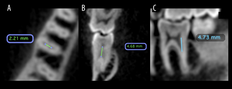
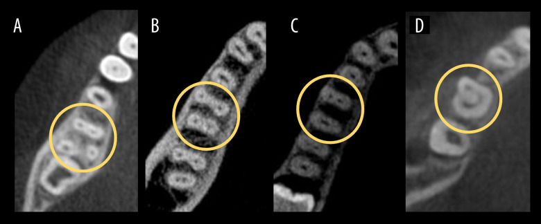
BACKGROUND The goal of conventional endodontic therapy is to clean the root canal system thoroughly, restore tooth function, and prevent re-infection. Success depends on understanding root canal morphology, resolving apical periodontitis, and using proper techniques. Studies highlight variations in root canal anatomy, with cone-beam computed tomography (CBCT) being crucial for accurate diagnosis despite its high cost and limited availability. MATERIAL AND METHODS This retrospective study reviewed CBCT images of 1820 (907 male, 913 female) patients aged 18 to 78 years. Analysis included 2081 mandibular first molars for variations in root and canal morphology and right- and left-side symmetry. Inter-orifice distance was measured, along with distance from the cementoenamel junction (CEJ) to the level of canal bifurcation. RESULTS In this study, 96.01% of teeth had 2 roots and 3.89% had 3 roots. The percentages of canal configuration were 77.70% for 3 canals, 21.58% for 4 canals, 0.67% for 2 canals, and 0.05% for 1 canal. The inter-orifice distance was 2.07 mm for 2 canals in 1 root and 2.86 mm for 2 canals in separate distal roots. Distance from the CEJ to canal bifurcation varied significantly between 2 canals within 1 distal root (3.35 mm), 2 canals in separate distal roots (1.60 mm), as well as between distal (3.35 mm) and mesial roots (1.10 mm). CONCLUSIONS In mandibular first molars, only 3.89% have additional distolingual roots. Sex and ethnicity showed no influence on number of roots and canals. Distal canals showed a deeper bifurcation and greater inter-orifice distance than did mesial canals.
传统牙髓治疗的目标是彻底清洁根管系统、恢复牙齿功能并预防再次感染。治疗成功与否取决于对根管形态的了解、根尖周炎的解决以及使用恰当的技术。研究强调根管解剖结构存在差异,尽管锥形束计算机断层扫描(CBCT)成本高且可用性有限,但对于准确诊断至关重要。
这项回顾性研究分析了1820名年龄在18至78岁之间患者(907名男性,913名女性)的CBCT图像。分析包括2081颗下颌第一磨牙的牙根和根管形态变化以及左右侧对称性。测量了根管口间距离以及从牙骨质釉质界(CEJ)到根管分叉水平的距离。
在本研究中,96.01%的牙齿有2个牙根,3.89%有3个牙根。根管形态的比例为:3根管占77.70%,4根管占21.58%,2根管占0.67%,1根管占0.05%。1个牙根内2根管的根管口间距离为2.07毫米,单独远中根内2根管的根管口间距离为2.86毫米。从CEJ到根管分叉的距离在1个远中根内的2根管(3.35毫米)、单独远中根内的2根管(1.60毫米)之间以及远中根(3.35毫米)和近中根(1.10毫米)之间有显著差异。
在下颌第一磨牙中,只有3.89%有额外的远舌根。性别和种族对牙根和根管数量没有影响。远中根管比近中根管显示出更深的分叉和更大的根管口间距离。