Section of Pulmonary and Critical Medicine, Department of Medicine, Lung Injury Center, University of Chicago, Chicago, Illinois, United States of America.
PLoS One. 2012;7(1):e30957. doi: 10.1371/journal.pone.0030957. Epub 2012 Jan 31.
Oxidation products of 1-palmitoyl-2-arachidonoyl-sn-glycero-3-phosphatidylcholine (OxPAPC) differentially modulate endothelial cell (EC) barrier function in a dose-dependent fashion. Vascular endothelial growth factor receptor-2 (VEGFR2) is involved in the OxPAPC-induced EC inflammatory activation. This study examined a role of VEGFR2 in barrier dysfunction caused by high concentrations of OxPAPC and evaluated downstream signaling mechanisms resulting from the effect of OxPAPC in EC from pulmonary and systemic circulation.
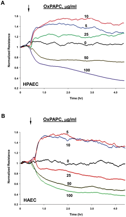
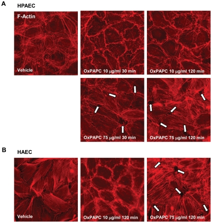
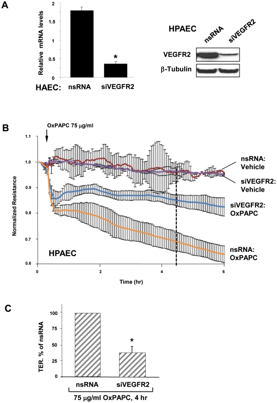
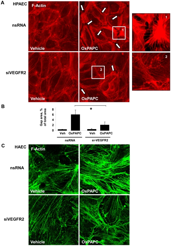
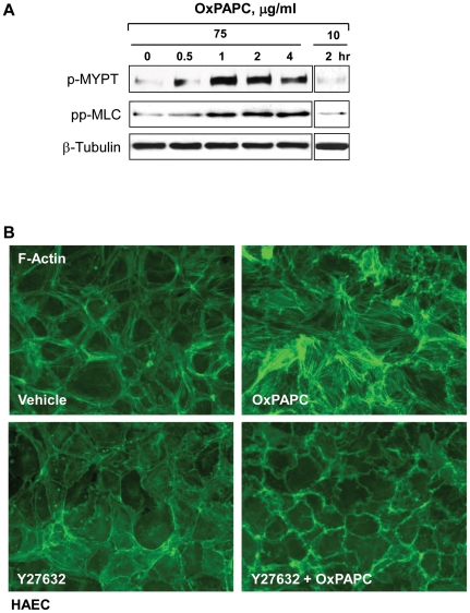
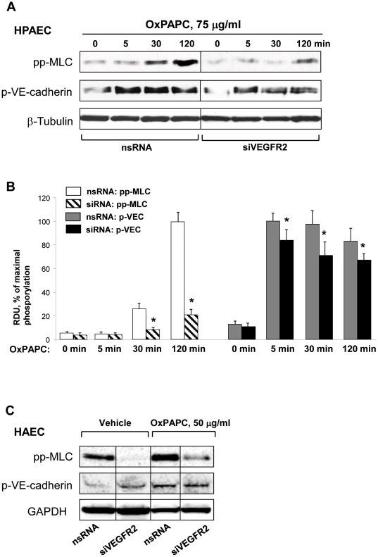
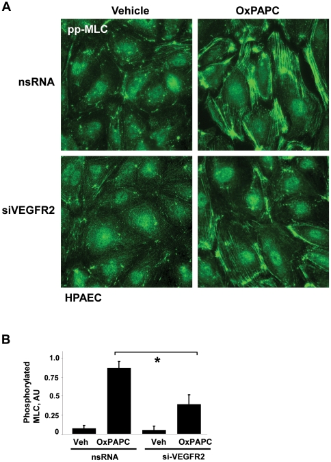
EC monolayer permeability in human pulmonary artery endothelial cells (HPAEC) and human aortic endothelial cells (HAEC) was monitored by changes in transendothelial electrical resistance (TER) across EC monolayers. Actin cytoskeleton was examined by immunostaining with Texas Red labeled phalloidin. Phosphorylation of myosin light chains (MLC) and VE-Cadherin was examined by Western blot and immunofluorescence techniques. The role of VEGFR2 in OxPAPC-induced permeability and cytoskeletal arrangement were determined using siRNA-induced VEGFR2 knockdown.
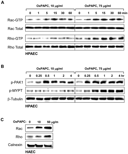
Low OxPAPC concentrations (5-20 µg/ml) induced a barrier protective response in both HPAEC and HAEC, while high OxPAPC concentrations (50-100 µg/ml) caused a rapid increase in permeability; actin stress fiber formation and increased MLC phosphorylation were observed as early as 30 min after treatment. VEGFR2 knockdown dramatically decreased the amount of MLC phosphorylation and stress fiber formation caused by high OxPAPC concentrations with modest effects on the amount of VE-cadherin phosphorylation at Y(731). We present evidence that activation of Rho is involved in the OxPAPC/VEGFR2 mechanism of EC permeability induced by high OxPAPC concentrations. Knockdown of VEGFR2 did not rescue the early drop in TER but prevented further development of OxPAPC-induced barrier dysfunction.
This study shows that VEGFR2 is involved in the delayed phase of EC barrier dysfunction caused by high OxPAPC concentrations and contributes to stress fiber formation and increased MLC phosphorylation.
1-棕榈酰基-2-花生四烯酰基-sn-甘油-3-磷酸胆碱(OxPAPC)的氧化产物以剂量依赖的方式差异化调节内皮细胞(EC)的屏障功能。血管内皮生长因子受体-2(VEGFR2)参与 OxPAPC 诱导的 EC 炎症激活。本研究探讨了 VEGFR2 在高浓度 OxPAPC 引起的屏障功能障碍中的作用,并评估了 OxPAPC 在肺循环和体循环内皮细胞中产生的下游信号机制。
通过测量跨内皮细胞单层的跨内皮电阻(TER)变化来监测人肺动脉内皮细胞(HPAEC)和人主动脉内皮细胞(HAEC)的 EC 单层通透性。通过用 Texas Red 标记的鬼笔环肽进行免疫染色来检查肌动蛋白细胞骨架。通过 Western blot 和免疫荧光技术检查肌球蛋白轻链(MLC)和 VE-钙粘蛋白的磷酸化。使用 siRNA 诱导的 VEGFR2 敲低来确定 VEGFR2 在 OxPAPC 诱导的通透性和细胞骨架排列中的作用。
低浓度 OxPAPC(5-20μg/ml)诱导 HPAEC 和 HAEC 产生屏障保护反应,而高浓度 OxPAPC(50-100μg/ml)则导致通透性迅速增加;在处理后 30 分钟即可观察到肌动蛋白应力纤维形成和 MLC 磷酸化增加。VEGFR2 敲低显著降低了高浓度 OxPAPC 引起的 MLC 磷酸化和应力纤维形成的量,对 Y(731)处 VE-钙粘蛋白磷酸化的量仅有适度影响。我们提供的证据表明,Rho 的激活参与了高浓度 OxPAPC/VEGFR2 引起的 EC 通透性机制。VEGFR2 敲低不能挽救早期 TER 的下降,但可防止 OxPAPC 诱导的屏障功能障碍的进一步发展。
本研究表明,VEGFR2 参与了高浓度 OxPAPC 引起的 EC 屏障功能障碍的迟发相,并有助于应力纤维形成和增加 MLC 磷酸化。