Oxford Centre for Functional MRI of the Brain (FMRIB), University of Oxford, Oxford, UK.
Neurorehabil Neural Repair. 2012 Jul-Aug;26(6):581-93. doi: 10.1177/1545968311433208. Epub 2012 Feb 9.
Failure of adaptive plasticity with increasing pathology is suggested to contribute to progression of disability in multiple sclerosis (MS). However, functional impairments can be reduced with practice, suggesting that brain plasticity is preserved even in patients with substantial damage.
. Here, functional magnetic resonance imaging (fMRI) was used to probe systems-level mechanisms of brain plasticity associated with improvements in visuomotor performance in MS patients and related to measures of microstructural damage.
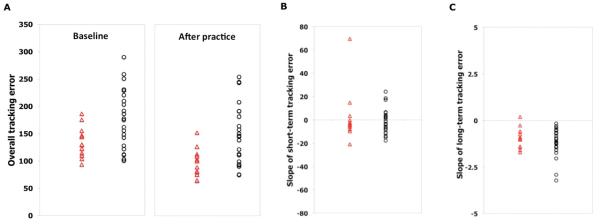
23 MS patients and 12 healthy controls underwent brain fMRI during the first practice session of a visuomotor task (short-term practice) and after 2 weeks of daily practice with the same task (longer-term practice). Participants also underwent a structural brain MRI scan.
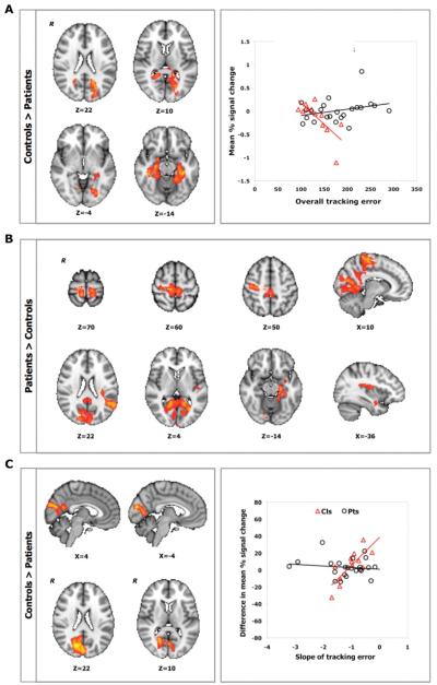
Patients performed more poorly than controls at baseline. Nonetheless, with practice, patients showed performance improvements similar to controls and independent of the extent of MRI measures of brain pathology. Different relationships between performance improvements and activations were found between groups: greater short-term improvements were associated with lower activation in the sensorimotor, posterior cingulate, and parahippocampal cortices for patients, whereas greater long-term improvements correlated with smaller activation reductions in the visual cortex of controls.
Brain plasticity for visuomotor practice is preserved in MS patients despite a high burden of cerebral pathology. Cognitive systems different from those acting in controls contribute to this plasticity in patients. These findings challenge the notion that increasing pathology is accompanied by an outright failure of adaptive plasticity, supporting a neuroscientific rationale for recovery-oriented strategies even in chronically disabled patients.
随着疾病的发展,适应性可塑性的失败被认为是导致多发性硬化症(MS)残疾进展的原因之一。然而,通过练习可以减轻功能障碍,这表明即使在有大量损伤的患者中,大脑的可塑性仍然得以保留。
本研究旨在使用功能磁共振成像(fMRI)来探究与 MS 患者的视动表现改善相关的大脑可塑性的系统水平机制,以及这些机制与脑微观结构损伤的测量指标之间的关系。
23 名 MS 患者和 12 名健康对照者在进行视动任务的第一次练习(短期练习)时,以及在相同任务的日常练习 2 周后(长期练习)进行了大脑 fMRI 扫描。参与者还接受了结构性脑 MRI 扫描。
患者在基线时的表现明显不如对照组。尽管如此,经过练习后,患者的表现改善与对照组相似,且与脑病理的 MRI 测量指标的严重程度无关。不同组之间的表现改善和激活之间存在不同的关系:对于患者而言,短期表现的改善与感觉运动、后扣带回和海马旁皮质的激活降低有关,而对于对照组而言,长期表现的改善与视觉皮层的激活减少程度较小有关。
尽管多发性硬化症患者的脑病理学负担很高,但他们对视动练习的大脑可塑性仍得以保留。与对照组不同的认知系统有助于患者的这种可塑性。这些发现挑战了随着病理学的增加,适应性可塑性会彻底失效的观点,为即使是慢性残疾患者也应采取以恢复为导向的策略提供了神经科学依据。