• 文献检索
  • 文档翻译
  • 深度研究
  • 学术资讯
  • Suppr Zotero 插件Zotero 插件
  • 邀请有礼
  • 套餐&价格
  • 历史记录
应用&插件
Suppr Zotero 插件Zotero 插件浏览器插件Mac 客户端Windows 客户端微信小程序
定价
高级版会员购买积分包购买API积分包
服务
文献检索文档翻译深度研究API 文档MCP 服务
关于我们
关于 Suppr公司介绍联系我们用户协议隐私条款
关注我们

Suppr 超能文献

核心技术专利:CN118964589B侵权必究
粤ICP备2023148730 号-1Suppr @ 2026

文献检索

告别复杂PubMed语法,用中文像聊天一样搜索,搜遍4000万医学文献。AI智能推荐,让科研检索更轻松。

立即免费搜索

文件翻译

保留排版,准确专业,支持PDF/Word/PPT等文件格式,支持 12+语言互译。

免费翻译文档

深度研究

AI帮你快速写综述,25分钟生成高质量综述,智能提取关键信息,辅助科研写作。

立即免费体验

用于评估肛瘘的磁共振成像

MRI for assessment of anal fistula.

作者信息

Torkzad Michael R, Karlbom Urban

出版信息

Insights Imaging. 2010 May;1(2):62-71. doi: 10.1007/s13244-010-0022-y. Epub 2010 May 27.

DOI:10.1007/s13244-010-0022-y
PMID:22347906
原文链接:https://pmc.ncbi.nlm.nih.gov/articles/PMC3259332/
Abstract

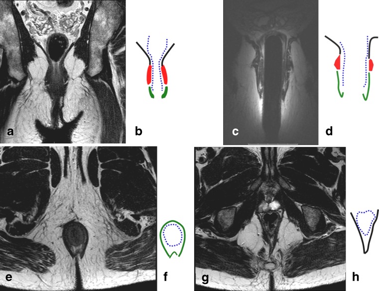
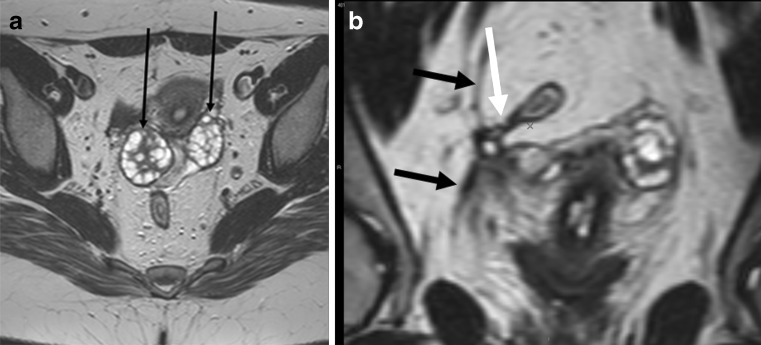
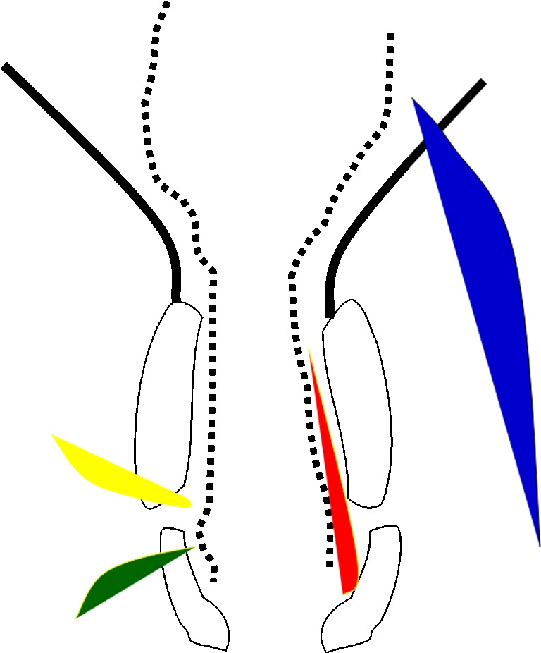
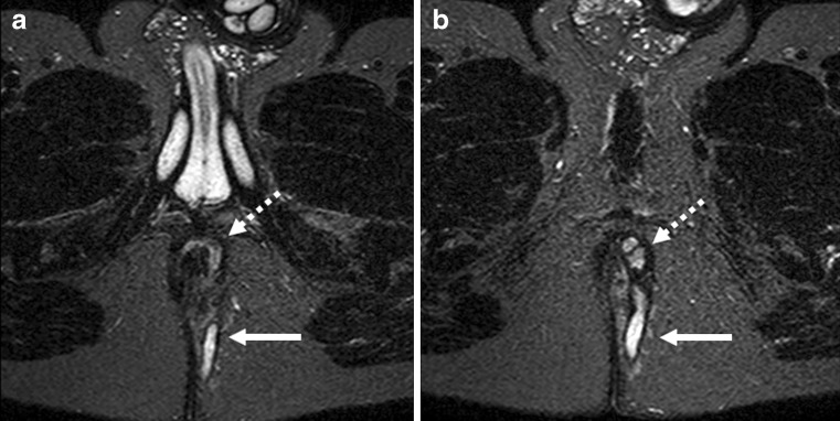
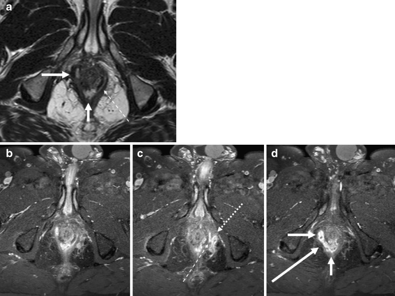
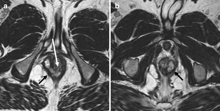
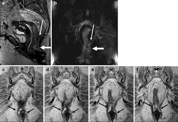
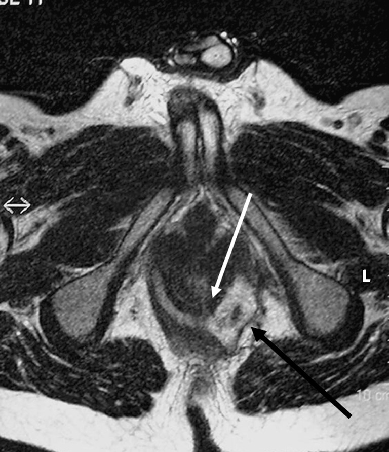
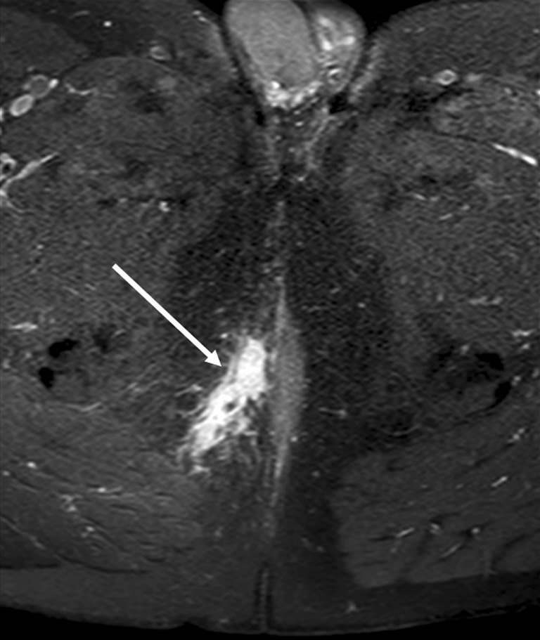
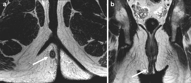
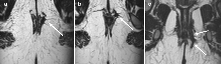
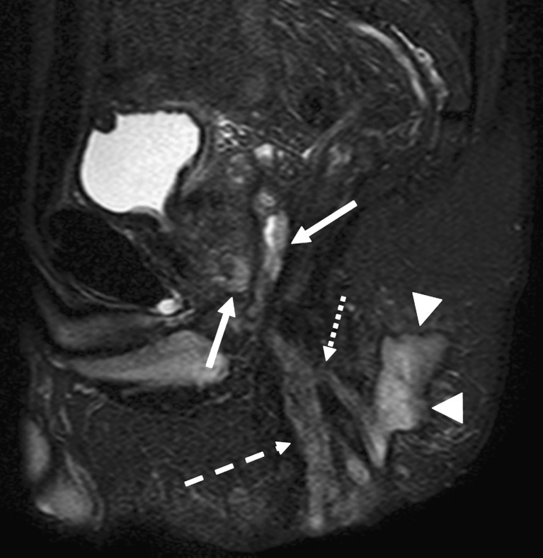
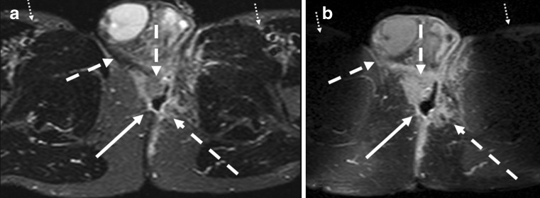
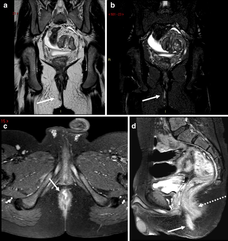
Magnetic resonance imaging (MRI) is the best imaging modality for preoperative assessment of patients with anal fistula. MRI helps to accurately demonstrate disease extension and predict prognosis. This in turn helps make therapy decisions and monitor therapy. The pertinent anatomy, fistula classification and MRI findings will be discussed.

摘要

磁共振成像(MRI)是肛瘘患者术前评估的最佳成像方式。MRI有助于准确显示疾病的蔓延情况并预测预后。这反过来有助于做出治疗决策并监测治疗效果。本文将讨论相关的解剖结构、肛瘘分类及MRI表现。

https://cdn.ncbi.nlm.nih.gov/pmc/blobs/79f0/3288838/dbe6a93756cc/13244_2010_22_Fig15_HTML.jpg
https://cdn.ncbi.nlm.nih.gov/pmc/blobs/79f0/3288838/0cc33a0584e7/13244_2010_22_Fig1_HTML.jpg
https://cdn.ncbi.nlm.nih.gov/pmc/blobs/79f0/3288838/1e3d3b09a334/13244_2010_22_Fig2_HTML.jpg
https://cdn.ncbi.nlm.nih.gov/pmc/blobs/79f0/3288838/b9b1e7449a7a/13244_2010_22_Fig3_HTML.jpg
https://cdn.ncbi.nlm.nih.gov/pmc/blobs/79f0/3288838/2a473975e424/13244_2010_22_Fig4_HTML.jpg
https://cdn.ncbi.nlm.nih.gov/pmc/blobs/79f0/3288838/0453378f210e/13244_2010_22_Fig5_HTML.jpg
https://cdn.ncbi.nlm.nih.gov/pmc/blobs/79f0/3288838/de8359ff5360/13244_2010_22_Fig6_HTML.jpg
https://cdn.ncbi.nlm.nih.gov/pmc/blobs/79f0/3288838/3edf7eec8478/13244_2010_22_Fig7_HTML.jpg
https://cdn.ncbi.nlm.nih.gov/pmc/blobs/79f0/3288838/8e7883e2614f/13244_2010_22_Fig8_HTML.jpg
https://cdn.ncbi.nlm.nih.gov/pmc/blobs/79f0/3288838/79197a188a40/13244_2010_22_Fig9_HTML.jpg
https://cdn.ncbi.nlm.nih.gov/pmc/blobs/79f0/3288838/a4ae82385e0b/13244_2010_22_Fig10_HTML.jpg
https://cdn.ncbi.nlm.nih.gov/pmc/blobs/79f0/3288838/516205edd356/13244_2010_22_Fig11_HTML.jpg
https://cdn.ncbi.nlm.nih.gov/pmc/blobs/79f0/3288838/cb2d8dd56955/13244_2010_22_Fig12_HTML.jpg
https://cdn.ncbi.nlm.nih.gov/pmc/blobs/79f0/3288838/0e0edd862d19/13244_2010_22_Fig13_HTML.jpg
https://cdn.ncbi.nlm.nih.gov/pmc/blobs/79f0/3288838/683be90cde77/13244_2010_22_Fig14_HTML.jpg
https://cdn.ncbi.nlm.nih.gov/pmc/blobs/79f0/3288838/dbe6a93756cc/13244_2010_22_Fig15_HTML.jpg

相似文献

1
MRI for assessment of anal fistula.用于评估肛瘘的磁共振成像
Insights Imaging. 2010 May;1(2):62-71. doi: 10.1007/s13244-010-0022-y. Epub 2010 May 27.
2
Evaluating postoperative anal fistula prognosis by diffusion-weighted MRI.应用弥散加权 MRI 评估术后肛瘘的预后。
Eur J Radiol. 2020 Nov;132:109294. doi: 10.1016/j.ejrad.2020.109294. Epub 2020 Sep 21.
3
The contribution of preoperative MRI to the surgical management of anal fistulas.术前磁共振成像在肛瘘手术治疗中的作用。
Diagn Interv Radiol. 2018 Nov;24(6):321-327. doi: 10.5152/dir.2018.18340.
4
Endoanal ultrasound or magnetic resonance imaging for preoperative assessment of anal fistula: a comparative study.经肛门超声或磁共振成像用于肛瘘术前评估的比较研究。
Colorectal Dis. 2001 May;3(3):189-97. doi: 10.1046/j.1463-1318.2001.00241.x.
5
[Application of magnetic resonance imaging in the diagnosis of complex anal fistula].[磁共振成像在复杂性肛瘘诊断中的应用]
Zhonghua Wei Chang Wai Ke Za Zhi. 2008 Jul;11(4):339-42.
6
A comparison of trans-fistula contrast-enhanced endoanal ultrasound and MRI in the diagnosis of anal fistula.经肛门瘘管对比增强腔内超声与 MRI 诊断肛瘘的比较。
Ann Palliat Med. 2021 Aug;10(8):9165-9173. doi: 10.21037/apm-21-1624.
7
Magnetic resonance imaging of perianal fistulas: predictive value for Parks classification and identification of the internal opening.磁共振成像在肛周瘘管中的应用:对 Parks 分类的预测价值及内口的定位。
Colorectal Dis. 2000 Nov 17;2(6):340-5. doi: 10.1046/j.1463-1318.2000.00181.x.
8
[Classification of anal fistulas based on magnetic resonance imaging].基于磁共振成像的肛瘘分类
Zhonghua Wei Chang Wai Ke Za Zhi. 2018 Dec 25;21(12):1391-1395.
9
MR imaging of fistula in ano: indications and contribution to surgical assessment.肛管瘘管的磁共振成像:适应证及对手术评估的作用
Acta Chir Belg. 2011 Nov-Dec;111(6):393-7. doi: 10.1080/00015458.2011.11680780.
10
A novel template for anorectal fistula reporting in anal endosonography and MRI - a practical concept.一种用于肛门直肠瘘在肛门内超声和磁共振成像中报告的新型模板——一个实用概念。
Med Ultrason. 2019 Nov 24;21(4):483-486. doi: 10.11152/mu-2154.

引用本文的文献

1
Magnetic resonance fistulography with percutaneous jelly: A novel and cost-effective technique.经皮注射凝胶的磁共振瘘管造影术:一种新颖且经济高效的技术。
SA J Radiol. 2025 Aug 15;29(1):3166. doi: 10.4102/sajr.v29i1.3166. eCollection 2025.
2
Clinical and Radiological Features of Perianal Fistula: An MRI-Based Study in Addis Ababa, Ethiopia.肛周瘘管的临床与放射学特征:埃塞俄比亚亚的斯亚贝巴基于磁共振成像的研究
Ethiop J Health Sci. 2024 Oct;34(Spec Iss 1):31-38. doi: 10.4314/ejhs.v34i1.6S.
3
Development and validation of the MRI-based deep learning classifier for distinguishing perianal fistulizing Crohn's disease from cryptoglandular fistula: a multicenter cohort study.

本文引用的文献

1
Imaging of perianal fistulas.肛周瘘管的影像学检查
Clin Gastroenterol Hepatol. 2009 Oct;7(10):1037-45. doi: 10.1016/j.cgh.2009.06.030. Epub 2009 Jul 31.
2
Perianal Crohn disease: evaluation of dynamic contrast-enhanced MR imaging as an indicator of disease activity.肛周克罗恩病:动态对比增强磁共振成像作为疾病活动指标的评估
Radiology. 2009 May;251(2):380-7. doi: 10.1148/radiol.2512072128.
3
Correlative anatomy of the anus and rectum.
Semin Ultrasound CT MR. 2008 Dec;29(6):400-8. doi: 10.1053/j.sult.2008.10.007.
基于磁共振成像的深度学习分类器用于鉴别肛周瘘管性克罗恩病与隐窝腺性瘘管的开发与验证:一项多中心队列研究
EClinicalMedicine. 2024 Nov 22;78:102940. doi: 10.1016/j.eclinm.2024.102940. eCollection 2024 Dec.
4
The role of MRI in perianal fistulizing disease: diagnostic imaging and classification systems to monitor disease activity.MRI在肛周瘘管性疾病中的作用:用于监测疾病活动的诊断成像和分类系统
Abdom Radiol (NY). 2025 Feb;50(2):589-597. doi: 10.1007/s00261-024-04455-w. Epub 2024 Aug 24.
5
Cellular angiofibroma arising from the rectocutaneous fistula in an adult: A case report.成人直肠皮肤瘘引发的细胞性血管纤维瘤:一例报告。
World J Clin Cases. 2024 Apr 6;12(10):1778-1784. doi: 10.12998/wjcc.v12.i10.1778.
6
Contemporary management of anorectal fistula.肛门直肠瘘的现代治疗
Surg Open Sci. 2024 Jan 2;17:40-43. doi: 10.1016/j.sopen.2023.12.005. eCollection 2024 Jan.
7
Preoperative magnetic resonance imaging of anal fistulas with scrotal extension: a retrospective study.合并阴囊延伸的肛瘘术前磁共振成像:一项回顾性研究
Front Surg. 2023 Jul 20;10:1224931. doi: 10.3389/fsurg.2023.1224931. eCollection 2023.
8
Evaluation of the Effectiveness of a Combination of Chinese Herbal Fumigation Sitz-Bath and Red Ointment in Managing Postoperative Wound Healing and Pain Control in Anal Fistula Patients.中药熏洗坐浴联合红油膏在肛瘘术后创面愈合及疼痛控制中的效果评价。
Contrast Media Mol Imaging. 2022 Sep 15;2022:1905279. doi: 10.1155/2022/1905279. eCollection 2022.
9
The development of a minimum dataset for MRI reporting of anorectal fistula: a multi-disciplinary, expert consensus process.肛门直肠瘘磁共振报告最小数据集的制定:多学科专家共识过程。
Eur Radiol. 2022 Dec;32(12):8306-8316. doi: 10.1007/s00330-022-08931-z. Epub 2022 Jun 23.
10
Congenital Cheek Fistula: A Rare Case of Developmental Abnormality in the Buccal Area.先天性颊瘘:颊部区域发育异常的罕见病例。
Cureus. 2021 Jun 15;13(6):e15657. doi: 10.7759/cureus.15657. eCollection 2021 Jun.
4
Pelvic MRI in children with Crohn disease and suspected perianal involvement.克罗恩病合并疑似肛周受累儿童的盆腔磁共振成像
Pediatr Radiol. 2007 Feb;37(2):201-8. doi: 10.1007/s00247-006-0372-2. Epub 2006 Dec 16.
5
Infliximab in fistulizing Crohn's disease.英夫利昔单抗治疗瘘管型克罗恩病
Gastroenterol Clin North Am. 2006 Dec;35(4):795-820. doi: 10.1016/j.gtc.2006.09.007.
6
Meta-analysis of randomized clinical trials comparing drainage alone vs primary sphincter-cutting procedures for anorectal abscess-fistula.比较单纯引流与一期括约肌切开术治疗肛管直肠脓肿-肛瘘的随机临床试验的荟萃分析。
Int J Colorectal Dis. 2006 Sep;21(6):602-9. doi: 10.1007/s00384-005-0060-y. Epub 2005 Nov 30.
7
Endoanal magnetic resonance imaging of fistula-in-ano: a comparison of STIR with gadolinium-enhanced techniques.肛管内肛瘘的磁共振成像:短T1反转恢复序列(STIR)与钆增强技术的比较
Acta Radiol. 2005 Feb;46(1):3-8. doi: 10.1080/02841850510015947.
8
Clinical examination, endosonography, and MR imaging in preoperative assessment of fistula in ano: comparison with outcome-based reference standard.临床检查、腔内超声检查及磁共振成像在肛管直肠瘘术前评估中的应用:与基于结果的参考标准比较
Radiology. 2004 Dec;233(3):674-81. doi: 10.1148/radiol.2333031724. Epub 2004 Oct 21.
9
Prospective comparison of hydrogen peroxide-enhanced three-dimensional endoanal ultrasonography and endoanal magnetic resonance imaging of perianal fistulas.过氧化氢增强型三维肛管超声检查与肛管磁共振成像对肛瘘的前瞻性比较
Dis Colon Rectum. 2003 Oct;46(10):1407-15. doi: 10.1007/s10350-004-6758-z.
10
MR imaging of fistula-in-ano.肛瘘的磁共振成像
Eur J Radiol. 2003 Aug;47(2):98-107. doi: 10.1016/s0720-048x(03)00162-1.