• 文献检索
  • 文档翻译
  • 深度研究
  • 学术资讯
  • Suppr Zotero 插件Zotero 插件
  • 邀请有礼
  • 套餐&价格
  • 历史记录
应用&插件
Suppr Zotero 插件Zotero 插件浏览器插件Mac 客户端Windows 客户端微信小程序
定价
高级版会员购买积分包购买API积分包
服务
文献检索文档翻译深度研究API 文档MCP 服务
关于我们
关于 Suppr公司介绍联系我们用户协议隐私条款
关注我们

Suppr 超能文献

核心技术专利:CN118964589B侵权必究
粤ICP备2023148730 号-1Suppr @ 2026

文献检索

告别复杂PubMed语法,用中文像聊天一样搜索,搜遍4000万医学文献。AI智能推荐,让科研检索更轻松。

立即免费搜索

文件翻译

保留排版,准确专业,支持PDF/Word/PPT等文件格式,支持 12+语言互译。

免费翻译文档

深度研究

AI帮你快速写综述,25分钟生成高质量综述,智能提取关键信息,辅助科研写作。

立即免费体验

髋关节磁共振关节造影:年轻患者的技术与影像表现谱

Magnetic resonance arthrography of the hip: technique and spectrum of findings in younger patients.

作者信息

Aubry Sébastien, Bélanger Danny, Giguère Caroline, Lavigne Martin

出版信息

Insights Imaging. 2010 May;1(2):72-82. doi: 10.1007/s13244-010-0023-x. Epub 2010 Jun 8.

DOI:10.1007/s13244-010-0023-x
PMID:22347907
原文链接:https://pmc.ncbi.nlm.nih.gov/articles/PMC3259340/
Abstract

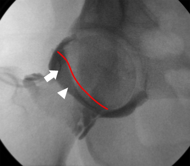
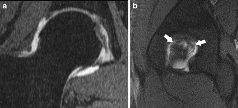
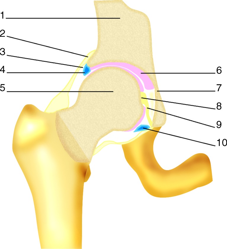
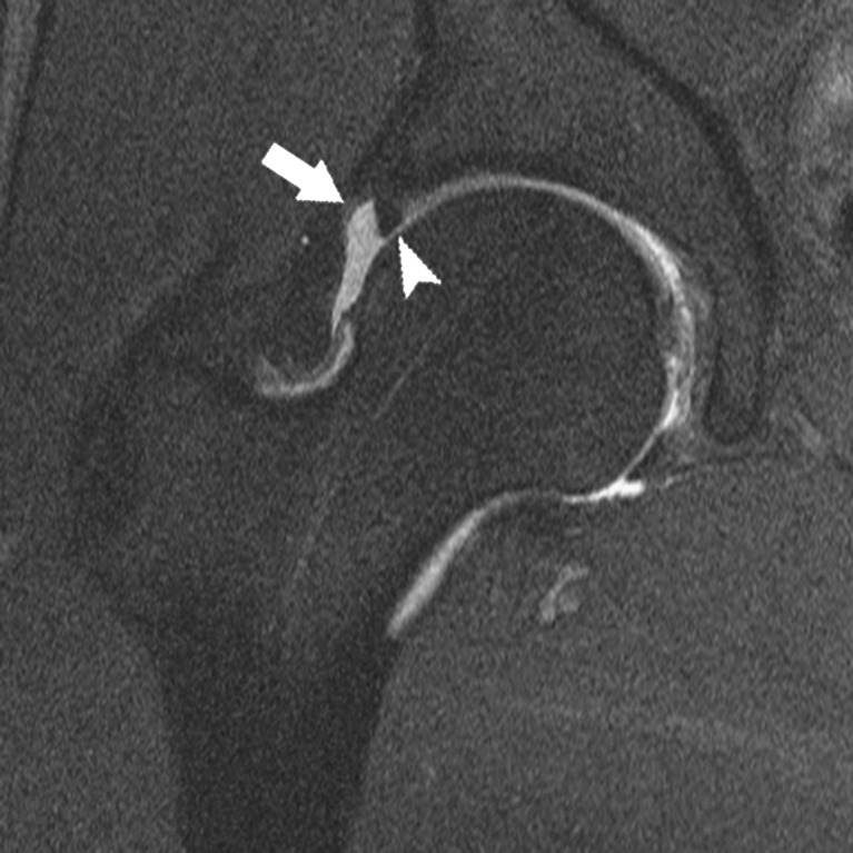
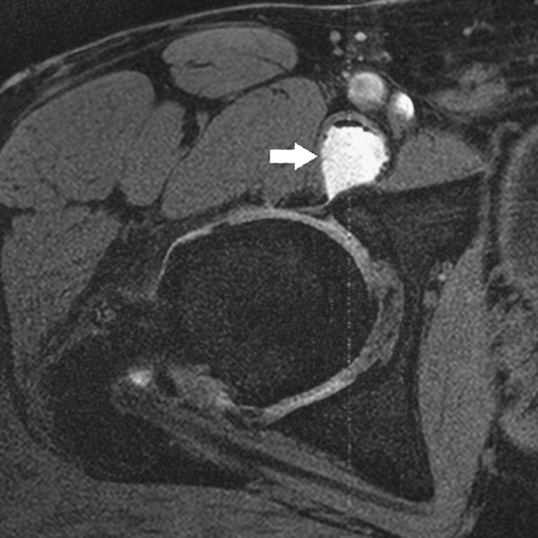
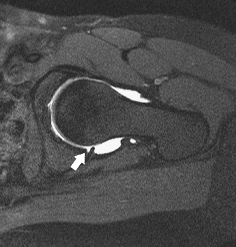
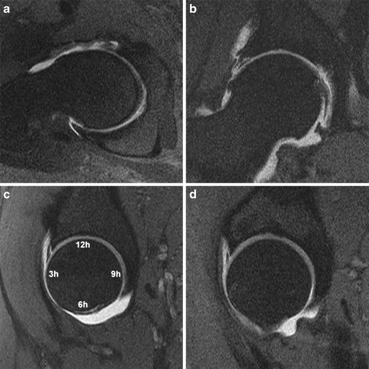
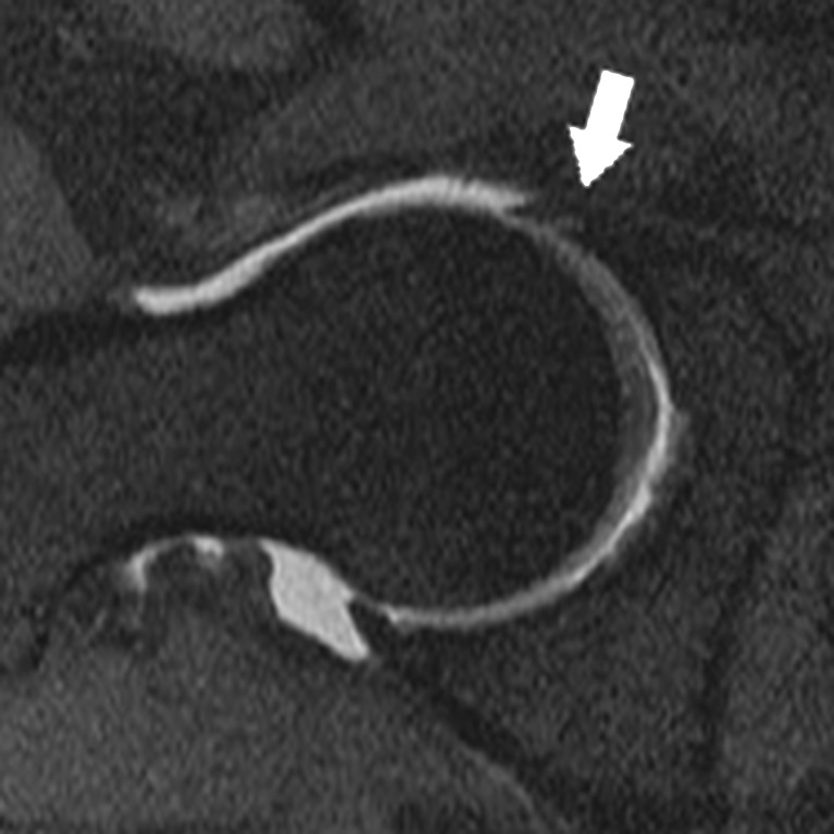
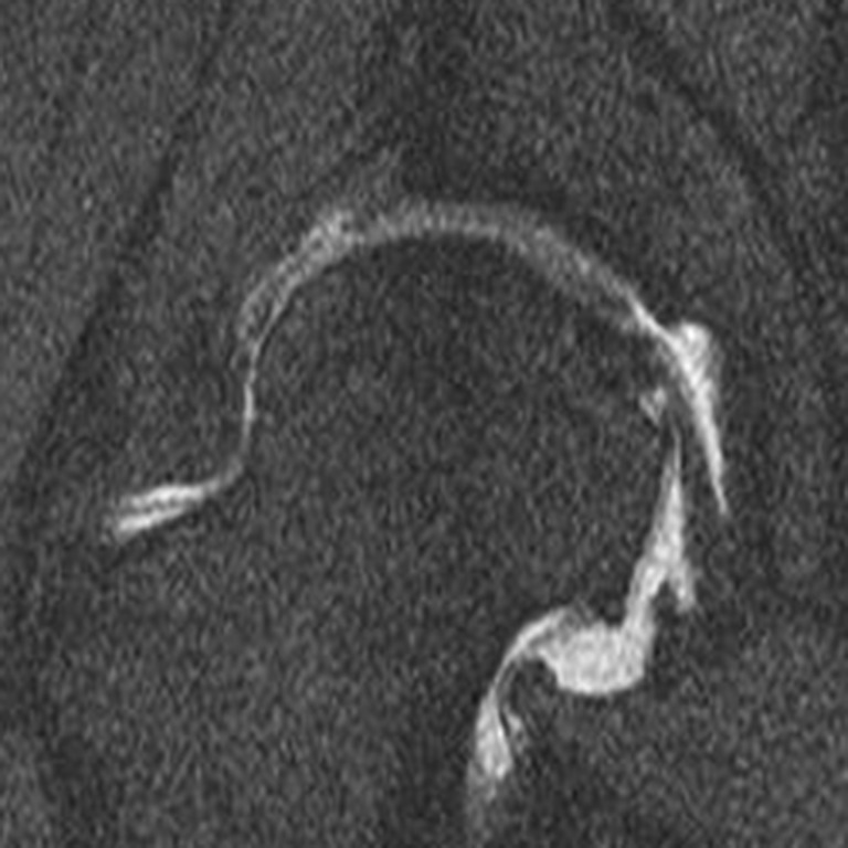
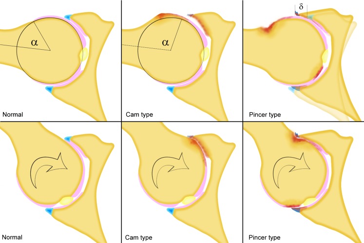
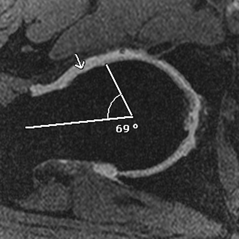
Magnetic resonance(MR) imaging is the reference imaging technique in the evaluation of hip abnormalities. However, in some pathological conditions-such as lesions of the labrum, cartilaginous lesions, femoroacetabular impingement, intra-articular foreign bodies, or in the pre-operative work-up of developmental dysplasia of the hip-intra-articular injection of a contrast medium is required to obtain a precise diagnosis. This article reviews the technical aspects, contraindications, normal appearance and potential pitfalls of MR arthrography, and illustrates the radiological appearance of commonly encountered conditions.

摘要

磁共振(MR)成像在评估髋关节异常方面是参考成像技术。然而,在某些病理情况下,如盂唇损伤、软骨损伤、股骨髋臼撞击症、关节内异物,或在髋关节发育不良的术前检查中,需要关节内注射造影剂以获得精确诊断。本文回顾了磁共振关节造影的技术方面、禁忌证、正常表现及潜在陷阱,并举例说明常见病症的影像学表现。

https://cdn.ncbi.nlm.nih.gov/pmc/blobs/f7d3/3288837/7e21bcec9d28/13244_2010_23_Fig15_HTML.jpg
https://cdn.ncbi.nlm.nih.gov/pmc/blobs/f7d3/3288837/d6513bb9ac41/13244_2010_23_Fig1_HTML.jpg
https://cdn.ncbi.nlm.nih.gov/pmc/blobs/f7d3/3288837/1add7a347ef2/13244_2010_23_Fig2_HTML.jpg
https://cdn.ncbi.nlm.nih.gov/pmc/blobs/f7d3/3288837/6eb476ba3e75/13244_2010_23_Fig3_HTML.jpg
https://cdn.ncbi.nlm.nih.gov/pmc/blobs/f7d3/3288837/0be3405066a4/13244_2010_23_Fig4_HTML.jpg
https://cdn.ncbi.nlm.nih.gov/pmc/blobs/f7d3/3288837/53018be48fd8/13244_2010_23_Fig5_HTML.jpg
https://cdn.ncbi.nlm.nih.gov/pmc/blobs/f7d3/3288837/0c97d1ad4ec6/13244_2010_23_Fig6_HTML.jpg
https://cdn.ncbi.nlm.nih.gov/pmc/blobs/f7d3/3288837/cbff98c93d5e/13244_2010_23_Fig7_HTML.jpg
https://cdn.ncbi.nlm.nih.gov/pmc/blobs/f7d3/3288837/c283da3082e7/13244_2010_23_Fig8_HTML.jpg
https://cdn.ncbi.nlm.nih.gov/pmc/blobs/f7d3/3288837/dbcf7c52ac79/13244_2010_23_Fig9_HTML.jpg
https://cdn.ncbi.nlm.nih.gov/pmc/blobs/f7d3/3288837/9b920f298c3d/13244_2010_23_Fig10_HTML.jpg
https://cdn.ncbi.nlm.nih.gov/pmc/blobs/f7d3/3288837/28b323e4e1d2/13244_2010_23_Fig11_HTML.jpg
https://cdn.ncbi.nlm.nih.gov/pmc/blobs/f7d3/3288837/b30b7a0c6b1b/13244_2010_23_Fig12_HTML.jpg
https://cdn.ncbi.nlm.nih.gov/pmc/blobs/f7d3/3288837/2ed915a312c6/13244_2010_23_Fig13_HTML.jpg
https://cdn.ncbi.nlm.nih.gov/pmc/blobs/f7d3/3288837/5a19d8991000/13244_2010_23_Fig14_HTML.jpg
https://cdn.ncbi.nlm.nih.gov/pmc/blobs/f7d3/3288837/7e21bcec9d28/13244_2010_23_Fig15_HTML.jpg

相似文献

1
Magnetic resonance arthrography of the hip: technique and spectrum of findings in younger patients.髋关节磁共振关节造影:年轻患者的技术与影像表现谱
Insights Imaging. 2010 May;1(2):72-82. doi: 10.1007/s13244-010-0023-x. Epub 2010 Jun 8.
2
Diagnosis of lesions of the acetabular labrum, of the labral-chondral transition zone, and of the cartilage in femoroacetabular impingement: Correlation between direct magnetic resonance arthrography and hip arthroscopy.髋臼盂唇、盂唇 - 软骨过渡区及股骨髋臼撞击症中软骨病变的诊断:直接磁共振关节造影与髋关节镜检查的相关性
Radiologia. 2015 Mar-Apr;57(2):131-41. doi: 10.1016/j.rx.2013.11.002. Epub 2014 Mar 19.
3
Hip MR arthrography and femoroacetabular impingement.髋关节磁共振关节造影与股骨髋臼撞击症
Semin Musculoskelet Radiol. 2006 Sep;10(3):208-19. doi: 10.1055/s-2006-957174.
4
[MR arthrography of the hip with emphasis on femoroacetabular impingement].[髋关节磁共振关节造影,重点关注股骨髋臼撞击症]
Radiologia. 2009 Jan-Feb;51(1):17-29; quiz 119. doi: 10.1016/S0033-8338(09)70402-3.
5
Evaluation of the acetabular labrum by MR arthrography.通过磁共振关节造影评估髋臼唇。
J Bone Joint Surg Br. 1997 Mar;79(2):230-4. doi: 10.1302/0301-620x.79b2.7288.
6
[Labral pathologies of the hip joint].[髋关节盂唇病变]
Radiologe. 2019 Mar;59(3):234-241. doi: 10.1007/s00117-018-0471-8.
7
[Magnetic resonance arthrography indications and technique (II). Lower limb].[磁共振关节造影的适应证与技术(II)。下肢]
Radiologia. 2006 Nov-Dec;48(6):357-68. doi: 10.1016/s0033-8338(06)75151-7.
8
MR Arthrography of the Hip.髋关节磁共振关节造影
Semin Musculoskelet Radiol. 1998;2(4):349-362. doi: 10.1055/s-2008-1080116.
9
Comparison Between 3-Dimensional Multiple-Echo Recombined Gradient Echo Magnetic Resonance Imaging and Arthroscopic Findings for the Evaluation of Acetabular Labrum Tear.三维多回波重组梯度回波磁共振成像与关节镜检查评估髋臼唇撕裂的比较。
Arthroscopy. 2019 Oct;35(10):2857-2865. doi: 10.1016/j.arthro.2019.05.006.
10
Imaging of the acetabular labrum.髋臼唇的影像学检查。
Semin Musculoskelet Radiol. 2013 Jul;17(3):248-57. doi: 10.1055/s-0033-1348091. Epub 2013 Jun 20.

引用本文的文献

1
A modified non-contrast MRI in the preoperative examination of periacetabular osteotomy at 3.0T.3.0T下改良非增强MRI在髋臼周围截骨术前检查中的应用
J Orthop Surg Res. 2025 Jan 31;20(1):121. doi: 10.1186/s13018-025-05521-9.
2
Utility of Magnetic Resonance Arthrography in the Age of Expanded Magnetic Resonance Imaging Capabilities: A Global Survey Perspective.在磁共振成像能力不断扩展的时代,磁共振关节造影的效用:一项全球调查视角
Cureus. 2024 Oct 15;16(10):e71526. doi: 10.7759/cureus.71526. eCollection 2024 Oct.
3
Magnetic Resonance Imaging and Magnetic Resonance Arthrography Are Both Reliable and Similar When Measuring Hip Capsule Thickness in Patients With Femoroacetabular Impingement Syndrome.

本文引用的文献

1
3 T magnetic resonance imaging of the musculoskeletal system.3T 磁共振成像在肌肉骨骼系统中的应用。
Radiol Med. 2010 Jun;115(4):571-84. doi: 10.1007/s11547-010-0521-4. Epub 2010 Feb 22.
2
Pain and other side effects after MR arthrography: prospective evaluation in 1085 patients.磁共振关节造影术后的疼痛及其他副作用:对1085例患者的前瞻性评估
Radiology. 2009 Mar;250(3):830-8. doi: 10.1148/radiol.2503080276. Epub 2009 Jan 21.
3
Comparison of MRI alpha angle measurement planes in femoroacetabular impingement.股骨髋臼撞击症中MRIα角测量平面的比较
在测量股骨髋臼撞击综合征患者的髋关节囊厚度时,磁共振成像和磁共振关节造影都可靠且相似。
Arthrosc Sports Med Rehabil. 2024 Feb 1;6(2):100874. doi: 10.1016/j.asmr.2023.100874. eCollection 2024 Apr.
4
SSR white paper: guidelines for utilization and performance of direct MR arthrography.SSR 白皮书:直接磁共振关节造影的使用和性能指南。
Skeletal Radiol. 2024 Feb;53(2):209-244. doi: 10.1007/s00256-023-04420-6. Epub 2023 Aug 11.
5
Image quality of hip MR arthrography with intra-articular injection of hyaluronic acid versus gadolinium-based contrast agent in patients with femoroacetabular impingement.髋关节 MRI 关节造影中关节内注射透明质酸与钆基对比剂在股骨髋臼撞击症患者中的图像质量比较。
Skeletal Radiol. 2020 Jun;49(6):937-944. doi: 10.1007/s00256-019-03366-y. Epub 2020 Jan 8.
6
Position paper on magnetic resonance imaging protocols in the musculoskeletal system (excluding the spine) by the Italian College of Musculoskeletal Radiology.意大利肌肉骨骼放射学会关于肌肉骨骼系统(不包括脊柱)磁共振成像协议的立场文件。
Radiol Med. 2019 Jun;124(6):522-538. doi: 10.1007/s11547-019-00992-3. Epub 2019 Jan 28.
Clin Orthop Relat Res. 2009 Mar;467(3):660-5. doi: 10.1007/s11999-008-0627-3. Epub 2008 Nov 27.
4
Impingement-free hip motion: the 'normal' angle alpha after osteochondroplasty.无撞击的髋关节运动:骨软骨成形术后的“正常”α角
Clin Orthop Relat Res. 2009 Mar;467(3):699-703. doi: 10.1007/s11999-008-0616-6. Epub 2008 Nov 19.
5
Direct MR arthrography of the hip with leg traction: feasibility for assessing articular cartilage.髋关节直接磁共振关节造影术结合腿部牵引:评估关节软骨的可行性
AJR Am J Roentgenol. 2008 Nov;191(5):W206; author reply W207. doi: 10.2214/AJR.08.1214.
6
MR arthrography of the hip: differentiation between an anterior sublabral recess as a normal variant and a labral tear.髋关节磁共振关节造影:区分正常变异的前盂唇下隐窝与盂唇撕裂。
Radiology. 2008 Dec;249(3):947-54. doi: 10.1148/radiol.2492080137. Epub 2008 Oct 7.
7
MR-guided direct arthrography of the hip.磁共振引导下的髋关节直接关节造影术。
J Magn Reson Imaging. 2008 Aug;28(2):462-5. doi: 10.1002/jmri.21432.
8
Direct MR arthrography of the hip with leg traction: feasibility for assessing articular cartilage.髋关节直接磁共振关节造影术结合腿部牵引:评估关节软骨的可行性
AJR Am J Roentgenol. 2008 Apr;190(4):1124-8. doi: 10.2214/AJR.07.2559.
9
Ischial spine projection into the pelvis : a new sign for acetabular retroversion.坐骨棘向骨盆内突出:髋臼后倾的一个新体征。
Clin Orthop Relat Res. 2008 Mar;466(3):677-83. doi: 10.1007/s11999-007-0058-6. Epub 2008 Feb 10.
10
The etiology of osteoarthritis of the hip: an integrated mechanical concept.髋关节骨关节炎的病因:一种综合力学概念。
Clin Orthop Relat Res. 2008 Feb;466(2):264-72. doi: 10.1007/s11999-007-0060-z. Epub 2008 Jan 10.