• 文献检索
  • 文档翻译
  • 深度研究
  • 学术资讯
  • Suppr Zotero 插件Zotero 插件
  • 邀请有礼
  • 套餐&价格
  • 历史记录
应用&插件
Suppr Zotero 插件Zotero 插件浏览器插件Mac 客户端Windows 客户端微信小程序
定价
高级版会员购买积分包购买API积分包
服务
文献检索文档翻译深度研究API 文档MCP 服务
关于我们
关于 Suppr公司介绍联系我们用户协议隐私条款
关注我们

Suppr 超能文献

核心技术专利:CN118964589B侵权必究
粤ICP备2023148730 号-1Suppr @ 2026

文献检索

告别复杂PubMed语法,用中文像聊天一样搜索,搜遍4000万医学文献。AI智能推荐,让科研检索更轻松。

立即免费搜索

文件翻译

保留排版,准确专业,支持PDF/Word/PPT等文件格式,支持 12+语言互译。

免费翻译文档

深度研究

AI帮你快速写综述,25分钟生成高质量综述,智能提取关键信息,辅助科研写作。

立即免费体验

微计算机断层扫描在体内诊断成像中的应用:肝/脑缺血和中风的啮齿动物模型的连续和对比评估。

In vivo diagnostic imaging using micro-CT: sequential and comparative evaluation of rodent models for hepatic/brain ischemia and stroke.

机构信息

Department of Anatomy and Neurobiology, Kinki University School of Medicine, Osaka-Sayama, Osaka, Japan.

出版信息

PLoS One. 2012;7(2):e32342. doi: 10.1371/journal.pone.0032342. Epub 2012 Feb 23.

DOI:10.1371/journal.pone.0032342
PMID:22384223
原文链接:https://pmc.ncbi.nlm.nih.gov/articles/PMC3285673/
Abstract

BACKGROUND

There is an increasing need for animal disease models for pathophysiological research and efficient drug screening. However, one of the technical barriers to the effective use of the models is the difficulty of non-invasive and sequential monitoring of the same animals. Micro-CT is a powerful tool for serial diagnostic imaging of animal models. However, soft tissue contrast resolution, particularly in the brain, is insufficient for detailed analysis, unlike the current applications of CT in the clinical arena. We address the soft tissue contrast resolution issue in this report.

METHODOLOGY

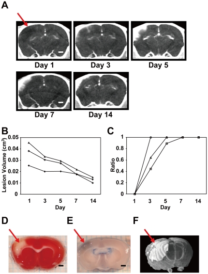
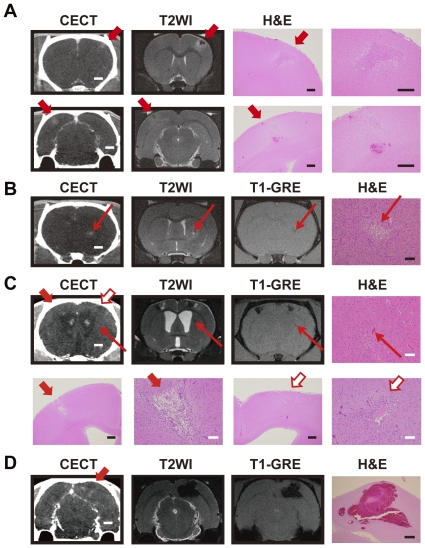
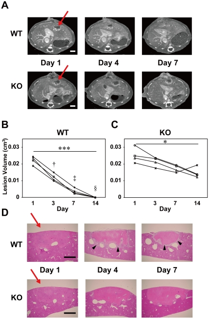
We performed contrast-enhanced CT (CECT) on mouse models of experimental cerebral infarction and hepatic ischemia. Pathological changes in each lesion were quantified for two weeks by measuring the lesion volume or the ratio of high attenuation area (%HAA), indicative of increased vascular permeability. We also compared brain images of stroke rats and ischemic mice acquired with micro-CT to those acquired with 11.7-T micro-MRI. Histopathological analysis was performed to confirm the diagnosis by CECT.

PRINCIPAL FINDINGS

In the models of cerebral infarction, vascular permeability was increased from three days through one week after surgical initiation, which was also confirmed by Evans blue dye leakage. Measurement of volume and %HAA of the liver lesions demonstrated differences in the recovery process between mice with distinct genetic backgrounds. Comparison of CT and MR images acquired from the same stroke rats or ischemic mice indicated that accuracy of volumetric measurement, as well as spatial and contrast resolutions of CT images, was comparable to that obtained with MRI. The imaging results were also consistent with the histological data.

CONCLUSIONS

This study demonstrates that the CECT scanning method is useful in rodents for both quantitative and qualitative evaluations of pathologic lesions in tissues/organs including the brain, and is also suitable for longitudinal observation of the same animals.

摘要

背景

对于病理生理学研究和高效药物筛选,动物疾病模型的需求日益增加。然而,有效使用这些模型的技术障碍之一是难以对同一动物进行非侵入性和连续监测。微计算机断层扫描(micro-CT)是对动物模型进行连续诊断成像的有力工具。然而,软组织对比分辨率,特别是在大脑中,对于详细分析来说还不够,这与 CT 在临床领域的当前应用不同。我们在本报告中解决了软组织对比分辨率问题。

方法

我们对实验性脑梗死和肝缺血的小鼠模型进行了对比增强 CT(CECT)检查。通过测量病变体积或高衰减区比例(%HAA),我们对每个病变的病理变化进行了两周的定量分析,这表明血管通透性增加。我们还比较了微 CT 采集的中风大鼠和缺血小鼠的脑图像与 11.7-T 微 MRI 采集的脑图像。通过 CECT 进行组织病理学分析以确认诊断。

主要发现

在脑梗死模型中,手术开始后三天至一周,血管通透性增加,伊文思蓝染料漏出也证实了这一点。肝病变的体积和%HAA 的测量表明,具有不同遗传背景的小鼠的恢复过程存在差异。来自同一中风大鼠或缺血小鼠的 CT 和 MR 图像的比较表明,体积测量的准确性以及 CT 图像的空间和对比分辨率与 MRI 相当。成像结果也与组织学数据一致。

结论

本研究表明,CECT 扫描方法在啮齿动物中对于包括大脑在内的组织/器官的病理病变的定量和定性评估都很有用,也适合对同一动物进行纵向观察。

https://cdn.ncbi.nlm.nih.gov/pmc/blobs/cc4b/3285673/f032ef1cc0f7/pone.0032342.g004.jpg
https://cdn.ncbi.nlm.nih.gov/pmc/blobs/cc4b/3285673/a473f7c54f4a/pone.0032342.g001.jpg
https://cdn.ncbi.nlm.nih.gov/pmc/blobs/cc4b/3285673/be5e46e9007e/pone.0032342.g002.jpg
https://cdn.ncbi.nlm.nih.gov/pmc/blobs/cc4b/3285673/dfc7ac05886a/pone.0032342.g003.jpg
https://cdn.ncbi.nlm.nih.gov/pmc/blobs/cc4b/3285673/f032ef1cc0f7/pone.0032342.g004.jpg

相似文献

1
In vivo diagnostic imaging using micro-CT: sequential and comparative evaluation of rodent models for hepatic/brain ischemia and stroke.微计算机断层扫描在体内诊断成像中的应用:肝/脑缺血和中风的啮齿动物模型的连续和对比评估。
PLoS One. 2012;7(2):e32342. doi: 10.1371/journal.pone.0032342. Epub 2012 Feb 23.
2
A new micro-computed tomography-based high-resolution blood-brain barrier imaging technique to study ischemic stroke.一种基于新的微计算机断层扫描的高分辨率血脑屏障成像技术,用于研究缺血性中风。
Stroke. 2014 Aug;45(8):2480-4. doi: 10.1161/STROKEAHA.114.006297. Epub 2014 Jul 10.
3
Rodent stroke induced by photochemical occlusion of proximal middle cerebral artery: evolution monitored with MR imaging and histopathology.大脑中动脉近端光化学闭塞诱导的啮齿动物中风:通过磁共振成像和组织病理学监测其演变
Eur J Radiol. 2007 Jul;63(1):68-75. doi: 10.1016/j.ejrad.2007.01.005. Epub 2007 Mar 6.
4
Nanoparticle contrast-enhanced micro-CT: A preclinical tool for the 3D imaging of liver and spleen in longitudinal mouse studies.纳米颗粒对比增强微型计算机断层扫描:一种用于小鼠纵向研究中肝脏和脾脏三维成像的临床前工具。
J Pharmacol Toxicol Methods. 2019 Mar-Apr;96:67-77. doi: 10.1016/j.vascn.2019.02.003. Epub 2019 Feb 7.
5
Validation of in vivo magnetic resonance imaging blood-brain barrier permeability measurements by comparison with gold standard histology.通过与金标准组织学比较验证体内磁共振成像血脑屏障通透性测量。
Stroke. 2011 Jul;42(7):2054-60. doi: 10.1161/STROKEAHA.110.597997. Epub 2011 Jun 2.
6
Micro-computed tomography in murine models of cerebral cavernous malformations as a paradigm for brain disease.微计算机断层扫描在脑海绵状血管畸形小鼠模型中作为脑部疾病的范例
J Neurosci Methods. 2016 Sep 15;271:14-24. doi: 10.1016/j.jneumeth.2016.06.021. Epub 2016 Jun 23.
7
A perfusion procedure for imaging of the mouse cerebral vasculature by X-ray micro-CT.一种通过 X 射线微计算机断层扫描对小鼠脑血管进行成像的灌注方法。
J Neurosci Methods. 2014 Jan 15;221:70-7. doi: 10.1016/j.jneumeth.2013.09.002. Epub 2013 Sep 19.
8
Implementation of dual- and triple-energy cone-beam micro-CT for postreconstruction material decomposition.双能和三能锥束显微CT在重建后材料分解中的应用。
Med Phys. 2008 Nov;35(11):5030-42. doi: 10.1118/1.2987668.
9
In vivo microCT imaging of rodent cerebral vasculature.啮齿动物脑血管的体内微型计算机断层扫描成像
Phys Med Biol. 2008 Apr 7;53(7):N99-107. doi: 10.1088/0031-9155/53/7/N01. Epub 2008 Mar 10.
10
Micro-CT based experimental liver imaging using a nanoparticulate contrast agent: a longitudinal study in mice.基于微 CT 的纳米颗粒造影剂实验性肝脏成像:小鼠的纵向研究。
PLoS One. 2011;6(9):e25692. doi: 10.1371/journal.pone.0025692. Epub 2011 Sep 30.

引用本文的文献

1
Turn-table micro-CT scanner for dynamic perfusion imaging in mice: design, implementation, and evaluation.用于小鼠动态灌注成像的转台式微型CT扫描仪:设计、实现与评估
Phys Med Biol. 2024 Aug 23;69(17). doi: 10.1088/1361-6560/ad6edd.
2
High-resolution hybrid micro-CT imaging pipeline for mouse brain region segmentation and volumetric morphometry.高分辨率混合微 CT 成像管道用于小鼠脑区分割和体积形态测量。
PLoS One. 2024 May 23;19(5):e0303288. doi: 10.1371/journal.pone.0303288. eCollection 2024.
3
Imaging bioluminescence by detecting localized haemodynamic contrast from photosensitized vasculature.

本文引用的文献

1
Evaluation of the middle cerebral artery occlusion techniques in the rat by in-vitro 3-dimensional micro- and nano computed tomography.利用体外三维微纳计算机断层扫描技术评估大鼠大脑中动脉闭塞模型。
BMC Neurol. 2010 May 28;10:36. doi: 10.1186/1471-2377-10-36.
2
Plasminogen is essential for granulation tissue formation during the recovery process after liver injury in mice.纤溶酶原对于小鼠肝损伤后恢复过程中的肉芽组织形成是必不可少的。
J Thromb Haemost. 2010 Jul;8(7):1555-66. doi: 10.1111/j.1538-7836.2010.03870.x. Epub 2010 Mar 23.
3
Initial brain lesion size affects the extent of subsequent pathophysiological responses.
通过检测光动力血管中的局部血流动力学对比来进行生物发光成像。
Nat Biomed Eng. 2024 Jun;8(6):775-786. doi: 10.1038/s41551-024-01210-w. Epub 2024 May 10.
4
An optimized CT-dense agent perfusion and micro-CT imaging protocol for chick embryo developmental stages.一种针对鸡胚发育阶段的优化CT造影剂灌注和微型CT成像方案。
BMC Biomed Eng. 2024 Apr 23;6(1):3. doi: 10.1186/s42490-024-00078-w.
5
High-resolution micro-CT for 3D infarct characterization and segmentation in mice stroke models.高分辨率微 CT 用于小鼠卒中模型的 3D 梗死特征分析和分割。
Sci Rep. 2022 Oct 19;12(1):17471. doi: 10.1038/s41598-022-21494-9.
6
Intra-arterial Contrast-enhanced Micro-computed Tomography Can Evaluate Intracranial Status in the Ultra-early Phase of Experimental Subarachnoid Hemorrhage in Rats.动脉内对比增强微计算机断层扫描可评估大鼠实验性蛛网膜下腔出血超早期的颅内状态。
Neurol Med Chir (Tokyo). 2021 Dec 15;61(12):721-730. doi: 10.2176/nmc.oa.2021-0027. Epub 2021 Oct 7.
7
Contrast enhanced X-ray computed tomography imaging of amyloid plaques in Alzheimer disease rat model on lab based micro CT system.基于实验室微 CT 系统的阿尔茨海默病大鼠模型中淀粉样斑块的对比增强 X 射线计算机断层成像。
Sci Rep. 2021 Mar 16;11(1):5999. doi: 10.1038/s41598-021-84579-x.
8
Reduction in Irradiation Dose in Aperture Coded Enhanced Computed Tomography Imager Using Super-Resolution Techniques.孔径编码增强计算机断层成像仪中利用超分辨率技术降低照射剂量。
Sensors (Basel). 2020 Nov 16;20(22):6551. doi: 10.3390/s20226551.
9
Stem Cell Repair of the Microvascular Damage in Stroke.干细胞修复脑卒中的微血管损伤。
Cells. 2020 Sep 11;9(9):2075. doi: 10.3390/cells9092075.
10
Neuroprotective Effects of Casein-Derived Peptide Met-Lys-Pro (MKP) in a Hypertensive Model.酪蛋白衍生肽Met-Lys-Pro(MKP)在高血压模型中的神经保护作用
Front Neurosci. 2020 Aug 14;14:845. doi: 10.3389/fnins.2020.00845. eCollection 2020.
初始脑损伤大小影响随后的病理生理反应的程度。
Brain Res. 2010 Mar 31;1322:109-17. doi: 10.1016/j.brainres.2010.01.077. Epub 2010 Feb 4.
4
Role of plasminogen in macrophage accumulation during liver repair.纤溶酶原在肝修复过程中巨噬细胞积累中的作用。
Thromb Res. 2010 May;125(5):e214-21. doi: 10.1016/j.thromres.2009.12.009. Epub 2010 Jan 10.
5
In vitro evaluation of the sinus sagittalis superior thrombosis model in the rat using 3D micro- and nanocomputed tomography.采用 3D 微纳计算机断层扫描技术对大鼠矢状窦血栓形成模型的体外评估。
Neuroradiology. 2010 Sep;52(9):815-21. doi: 10.1007/s00234-009-0617-5. Epub 2009 Nov 18.
6
Vascular imaging in small rodents using micro-CT.小动物的血管成像采用 micro-CT。
Methods. 2010 Jan;50(1):26-35. doi: 10.1016/j.ymeth.2009.09.003. Epub 2009 Sep 20.
7
Application of micro-CT in small animal imaging.微计算机断层扫描在小动物成像中的应用。
Methods. 2010 Jan;50(1):2-13. doi: 10.1016/j.ymeth.2009.08.007. Epub 2009 Aug 23.
8
Immunohistochemical analysis of brain lesions using S100B and glial fibrillary acidic protein antibodies in arundic acid- (ONO-2506) treated stroke-prone spontaneously hypertensive rats.用 S100B 和胶质纤维酸性蛋白抗体对尿酸(ONO-2506)处理的易卒中型自发性高血压大鼠脑损伤进行免疫组织化学分析。
J Neural Transm (Vienna). 2009 Oct;116(10):1209-19. doi: 10.1007/s00702-009-0278-x. Epub 2009 Aug 6.
9
In vivo micro-CT imaging of rat brain glioma: a comparison with 3T MRI and histology.大鼠脑胶质瘤的体内微型计算机断层扫描成像:与3T磁共振成像及组织学的比较
Neurosci Lett. 2009 Jul 10;458(1):28-31. doi: 10.1016/j.neulet.2009.04.033. Epub 2009 Apr 18.
10
Noninvasive monitoring of a murine model of metastatic pheochromocytoma: a comparison of contrast-enhanced microCT and nonenhanced MRI.转移性嗜铬细胞瘤小鼠模型的无创监测:对比增强微型计算机断层扫描与非增强磁共振成像的比较
J Magn Reson Imaging. 2009 Mar;29(3):685-91. doi: 10.1002/jmri.21654.