Department of Orthopaedics, The Third Affiliated Hospital, Sun Yat-sen University, Guangzhou, China.
PLoS One. 2012;7(2):e32356. doi: 10.1371/journal.pone.0032356. Epub 2012 Feb 27.
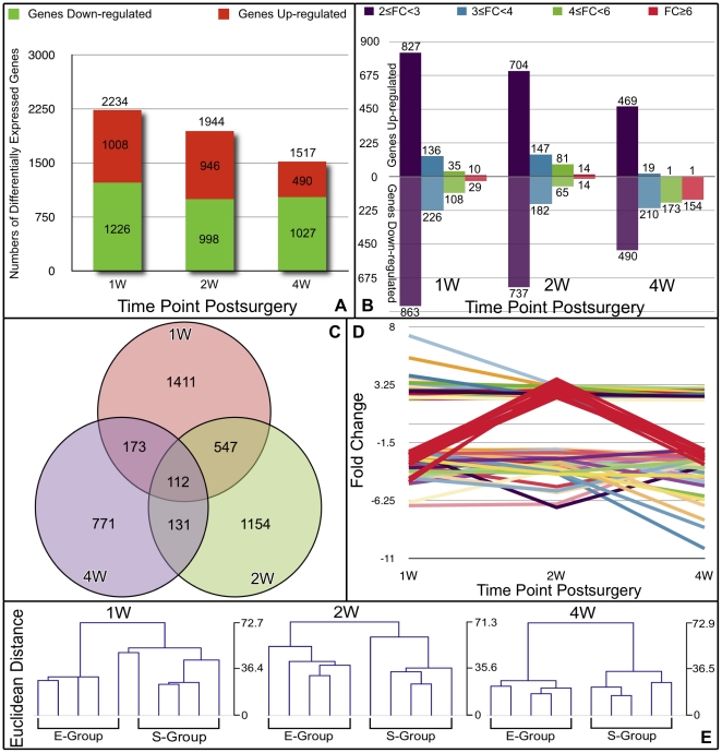
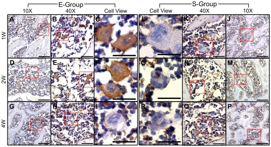
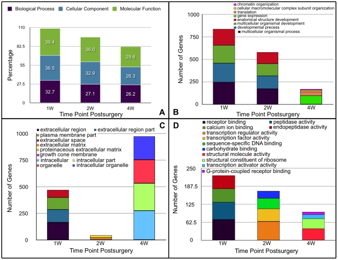
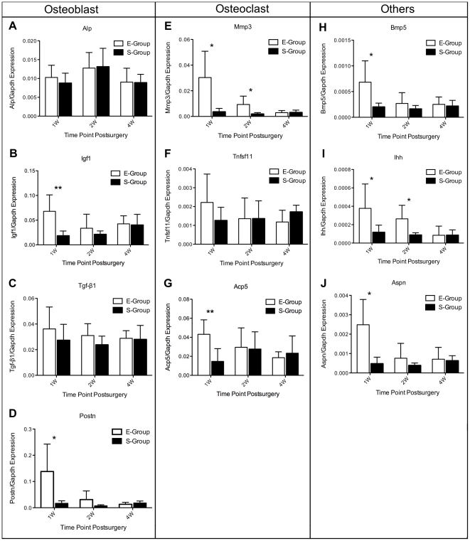
Osteoarthritis (OA) is a degenerative joint disease that affects both cartilage and bone. A better understanding of the early molecular changes in subchondral bone may help elucidate the pathogenesis of OA. We used microarray technology to investigate the time course of molecular changes in the subchondral bone in the early stages of experimental osteoarthritis in a rat model. We identified 2,234 differentially expressed (DE) genes at 1 week, 1,944 at 2 weeks and 1,517 at 4 weeks post-surgery. Further analyses of the dysregulated genes indicated that the events underlying subchondral bone remodeling occurred sequentially and in a time-dependent manner at the gene expression level. Some of the identified dysregulated genes that were identified have suspected roles in bone development or remodeling; these genes include Alp, Igf1, Tgf β1, Postn, Mmp3, Tnfsf11, Acp5, Bmp5, Aspn and Ihh. The differences in the expression of these genes were confirmed by real-time PCR, and the results indicated that our microarray data accurately reflected gene expression patterns characteristic of early OA. To validate the results of our microarray analysis at the protein level, immunohistochemistry staining was used to investigate the expression of Mmp3 and Aspn protein in tissue sections. These analyses indicate that Mmp3 protein expression completely matched the results of both the microarray and real-time PCR analyses; however, Aspn protein expression was not observed to differ at any time. In summary, our study demonstrated a simple method of separation of subchondral bone sample from the knee joint of rat, which can effectively avoid bone RNA degradation. These findings also revealed the gene expression profiles of subchondral bone in the rat OA model at multiple time points post-surgery and identified important DE genes with known or suspected roles in bone development or remodeling. These genes may be novel diagnostic markers or therapeutic targets for OA.
骨关节炎(OA)是一种影响软骨和骨骼的退行性关节疾病。更好地了解软骨下骨的早期分子变化可能有助于阐明 OA 的发病机制。我们使用微阵列技术研究了大鼠 OA 模型中软骨下骨在早期阶段的分子变化的时间进程。我们在手术后 1 周、2 周和 4 周时分别发现了 2234 个差异表达(DE)基因、1944 个和 1517 个。对失调基因的进一步分析表明,软骨下骨重塑的潜在事件在基因表达水平上依次且具有时间依赖性地发生。一些被鉴定出的失调基因被怀疑在骨骼发育或重塑中发挥作用;这些基因包括 Alp、Igf1、Tgfβ1、Postn、Mmp3、Tnfsf11、Acp5、Bmp5、Aspn 和 Ihh。这些基因的表达差异通过实时 PCR 得到了证实,结果表明我们的微阵列数据准确地反映了早期 OA 的基因表达模式。为了验证微阵列分析的结果,我们使用免疫组织化学染色来研究组织切片中 Mmp3 和 Aspn 蛋白的表达。这些分析表明,Mmp3 蛋白表达完全与微阵列和实时 PCR 分析的结果相匹配;然而,在任何时间点都没有观察到 Aspn 蛋白表达的差异。总之,我们的研究提供了一种从大鼠膝关节分离软骨下骨样本的简单方法,可以有效地避免骨 RNA 降解。这些发现还揭示了术后多个时间点大鼠 OA 模型软骨下骨的基因表达谱,并确定了具有已知或疑似在骨骼发育或重塑中发挥作用的重要 DE 基因。这些基因可能是 OA 的新型诊断标志物或治疗靶点。