National Laboratory of Biomacromolecules, Institute of Biophysics, Chinese Academy of Sciences, Beijing 100101, China.
J Cell Biol. 2012 Apr 2;197(1):27-35. doi: 10.1083/jcb.201111053. Epub 2012 Mar 26.
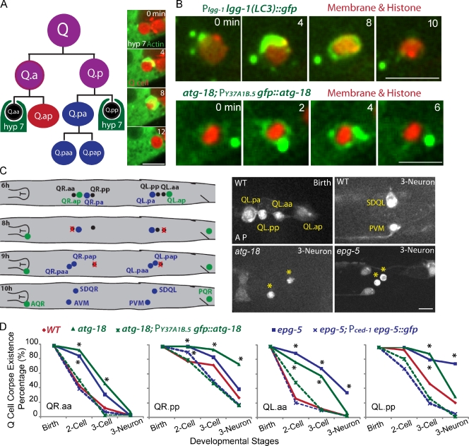
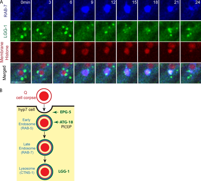
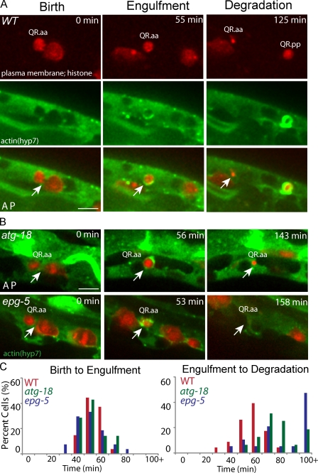
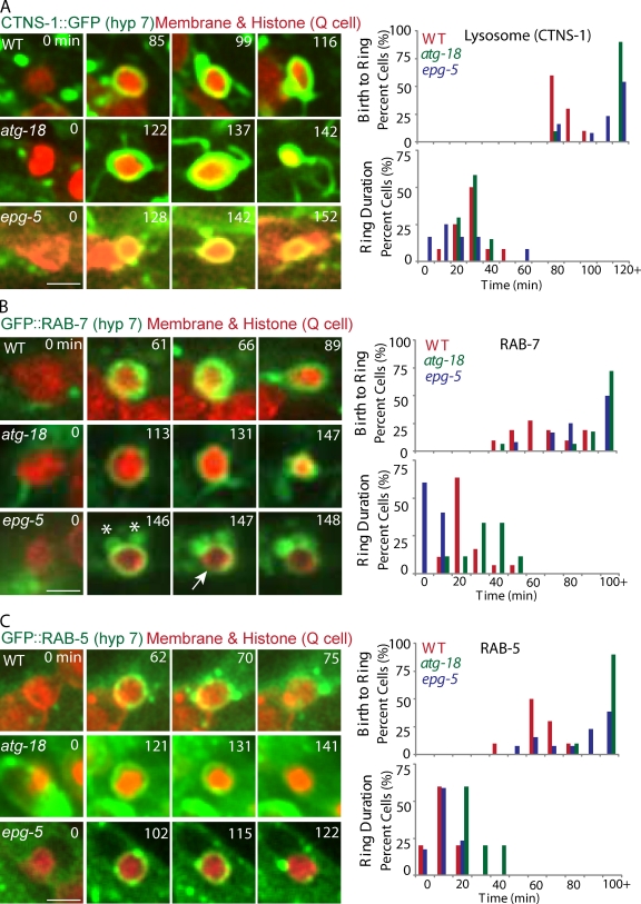
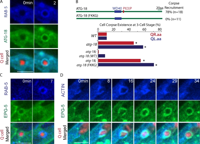
Apoptotic cell degradation is a fundamental process for organism development, and impaired clearance causes inflammatory or autoimmune disease. Although autophagy genes were reported to be essential for exposing the engulfment signal on apoptotic cells, their roles in phagocytes for apoptotic cell removal are not well understood. In this paper, we develop live-cell imaging techniques to study apoptotic cell clearance in the Caenorhabditis elegans Q neuroblast lineage. We show that the autophagy proteins LGG-1/LC3, ATG-18, and EPG-5 were sequentially recruited to internalized apoptotic Q cells in the phagocyte. In atg-18 or epg-5 mutants, apoptotic Q cells were internalized but not properly degraded; this phenotype was fully rescued by the expression of autophagy genes in the phagocyte. Time-lapse analysis of autophagy mutants revealed that recruitment of the small guanosine triphosphatases RAB-5 and RAB-7 to the phagosome and the formation of phagolysosome were all significantly delayed. Thus, autophagy genes act within the phagocyte to promote apoptotic cell degradation.
细胞凋亡的降解是生物体发育的一个基本过程,而清除功能受损会导致炎症或自身免疫性疾病。虽然自噬基因被报道对于在凋亡细胞上暴露吞噬信号是必需的,但它们在吞噬细胞中对于凋亡细胞清除的作用还不是很清楚。在本文中,我们开发了活细胞成像技术来研究秀丽隐杆线虫 Q 神经母细胞谱系中的凋亡细胞清除。我们表明,自噬蛋白 LGG-1/LC3、ATG-18 和 EPG-5 被顺序募集到吞噬细胞内的内化凋亡 Q 细胞。在 atg-18 或 epg-5 突变体中,凋亡 Q 细胞被内化但不能被适当降解;这种表型可以通过在吞噬细胞中表达自噬基因得到完全挽救。自噬突变体的延时分析显示,小 GTPase RAB-5 和 RAB-7 向吞噬体的募集以及吞噬溶酶体的形成都显著延迟。因此,自噬基因在吞噬细胞内发挥作用,促进凋亡细胞的降解。