Section of Human Anatomy, Faculty of Pharmacy, University G. d’Annunzio Chieti-Pescara, via dei Vestini 31, 66100 Chieti, Italy.
Eur J Histochem. 2012 Jan 31;56(1):e2. doi: 10.4081/ejh.2012.e2.
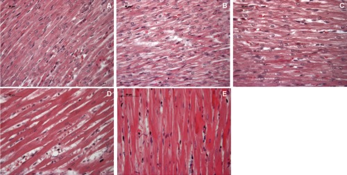
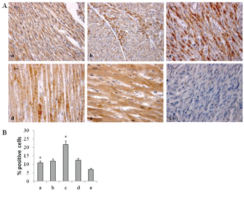
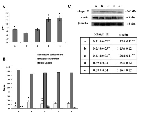
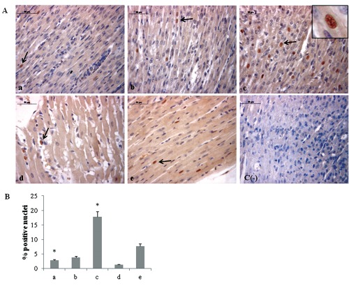
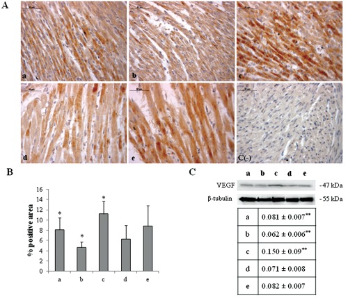
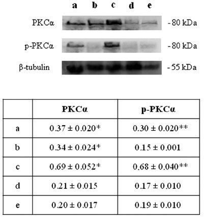
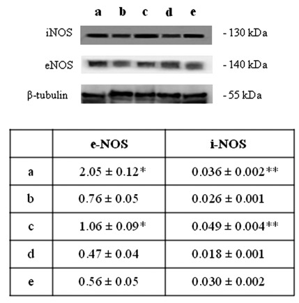
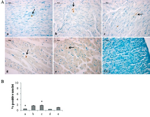
In premature babies birth an high oxygen level exposure can occur and newborn hyperoxia exposure can be associated with free radical oxygen release with impairment of myocardial function, while in adult animal models short exposure to hyperoxia seems to protect heart against ischemic injury. Thus, the mechanisms and consequences which take place after hyperoxia exposure are different and related to animals age. The aim of our work has been to analyze the role played by HIF-1α in the occurrence of the morphological modifications upon hyperoxia exposure in neonatal rat heart. Hyperoxia exposure induces connective compartment increase which seems to allow enhanced blood vessels growth. An increased hypoxia inducible factor-1α (HIF-1α) translocation and vascular endothelial growth factor (VEGF) expression has been found upon 95% oxygen exposure to induce morphological modifications. Upstream pPKC-α expression increase in newborn rats exposed to 95% oxygen can suggest PKC involvement in HIF-1α activation. Since nitric oxide synthase (NOS) are involved in heart vascular regulation, endothelial NOS (e-NOS) and inducible NOS (i-NOS) expression has been investigated: a lower eNOS and an higher iNOS expression has been found in newborn rats exposed to 95% oxygen related to the evidence that hyperoxia provokes a systemic vasoconstriction and to the iNOS pro-apoptotic action, respectively. The occurrence of apoptotic events, evaluated by TUNEL and Bax expression analyses, seems more evident in sample exposed to severe hyperoxia. All in all such results suggest that in newborn rats hyperoxia can trigger oxygen free radical mediated membrane injury through a pPKCα mediated HIF-1α signalling system, even though specificity of such response could be obtained by in vivo administration to the rats of specific inhibitors of PKCα. This intracellular signalling can switch molecular events leading to blood vessels development in parallel to pro-apoptotic events due to an immature anti-oxidant defensive system in newborn rat hearts.
在早产儿中,出生时可能会暴露于高氧环境中,而新生儿的高氧暴露可能与自由基氧释放有关,从而损害心肌功能,而在成年动物模型中,短暂暴露于高氧似乎可以保护心脏免受缺血性损伤。因此,高氧暴露后发生的机制和后果因动物年龄而异。我们的工作旨在分析 HIF-1α 在新生大鼠心脏高氧暴露后形态改变中的作用。高氧暴露诱导结缔组织间隙增加,似乎允许增强血管生长。我们发现,在 95%氧气暴露下,缺氧诱导因子-1α(HIF-1α)易位和血管内皮生长因子(VEGF)表达增加,从而导致形态改变。在新生大鼠中,95%氧气暴露时上游 pPKC-α表达增加,这表明 PKC 参与了 HIF-1α 的激活。由于一氧化氮合酶(NOS)参与心脏血管调节,因此研究了内皮型 NOS(e-NOS)和诱导型 NOS(i-NOS)的表达:在暴露于 95%氧气的新生大鼠中,eNOS 表达降低,iNOS 表达升高,这与高氧引起全身血管收缩和 iNOS 促凋亡作用有关。通过 TUNEL 和 Bax 表达分析评估的凋亡事件的发生似乎在暴露于严重高氧的样本中更为明显。总之,这些结果表明,在新生大鼠中,高氧可以通过 pPKCα 介导的 HIF-1α 信号系统触发氧自由基介导的膜损伤,尽管通过向大鼠体内给予 PKCα 的特异性抑制剂可以获得这种反应的特异性。这种细胞内信号可以切换分子事件,导致血管发育,同时由于新生大鼠心脏不成熟的抗氧化防御系统,导致促凋亡事件。