Department of Brain Repair and Rehabilitation, UCL Institute of Neurology, London WC1N 3BG, UK.
J Neurol Neurosurg Psychiatry. 2012 Jun;83(6):629-37. doi: 10.1136/jnnp-2011-301875. Epub 2012 Apr 6.
Traumatic spinal cord injury (SCI) leads to disruption of axonal architecture and macroscopic tissue loss with impaired information flow between the brain and spinal cord-the presumed basis of ensuing clinical impairment.
The authors used a clinically viable, multimodal MRI protocol to quantify the axonal integrity of the cranial corticospinal tract (CST) and to establish how microstructural white matter changes in the CST are related to cross-sectional spinal cord area and cortical reorganisation of the sensorimotor system in subjects with traumatic SCI.
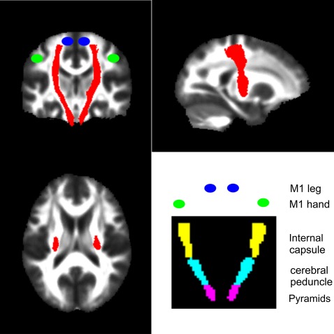
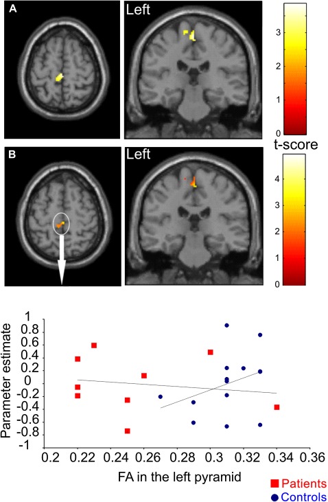
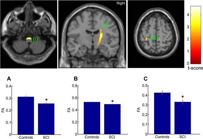
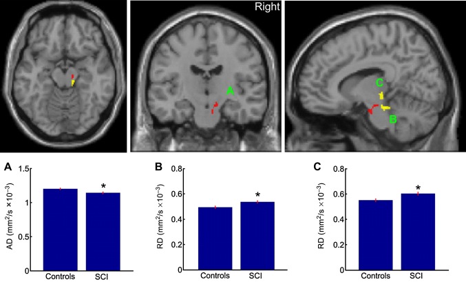
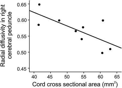
Nine volunteers with cervical injuries resulting in bilateral motor impairment and 14 control subjects were studied. The authors used diffusion tensor imaging to assess white matter integrity in the CST, T1-weighted imaging to measure cross-sectional spinal cord area and functional MRI to compare motor task-related brain activations. The relationships among microstructural, macrostructural and functional measures were assessed using regression analyses. Results Diffusion tensor imaging revealed significant differences in the CST of SCI subjects-compared with controls-in the pyramids, the internal capsule, the cerebral peduncle and the hand area. The microstructural white matter changes observed in the left pyramid predicted increased task-related responses in the left M1 leg area, while changes in the cerebral peduncle were predicted by reduced cord area.
The observed microstructural changes suggest trauma-related axonal degeneration and demyelination, which are related to cortical motor reorganisation and macrostructure. The extent of these changes may reflect the plasticity of motor pathways associated with cortical reorganisation. This clinically viable multimodal imaging approach is therefore appropriate for monitoring degeneration of central pathways and the evaluation of treatments targeting axonal repair in SCI.
外伤性脊髓损伤(SCI)导致轴突结构中断和宏观组织丢失,大脑和脊髓之间的信息传递受损——这被认为是随后临床损伤的基础。
作者使用一种可行的多模态 MRI 方案来量化颅皮质脊髓束(CST)的轴突完整性,并确定 CST 的微观结构白质变化与外伤性 SCI 患者的脊髓横断面面积和感觉运动系统皮质重组之间的关系。
研究了 9 名因颈损伤导致双侧运动障碍的志愿者和 14 名对照受试者。作者使用弥散张量成像来评估 CST 的白质完整性,使用 T1 加权成像来测量脊髓横断面面积,并使用功能磁共振成像来比较运动任务相关的大脑激活。使用回归分析评估微观结构、宏观结构和功能测量之间的关系。结果弥散张量成像显示,SCI 受试者的 CST 在锥体、内囊、大脑脚和手部区域与对照组存在显著差异。左侧锥体观察到的微观结构白质变化预测了左侧 M1 腿部区域的任务相关反应增加,而大脑脚的变化则由脊髓面积减少预测。
观察到的微观结构变化提示与创伤相关的轴突变性和脱髓鞘,这与皮质运动重组和宏观结构有关。这些变化的程度可能反映了与皮质重组相关的运动通路的可塑性。因此,这种可行的临床多模态成像方法适用于监测中枢通路的退化和评估针对 SCI 轴突修复的治疗效果。