Department of Radiology, Chungnam National University Hospital, Daejeon, Korea.
J Breast Cancer. 2012 Mar;15(1):57-64. doi: 10.4048/jbc.2012.15.1.57. Epub 2012 Mar 28.
The purpose of this study is to evaluate imaging and histopathologic findings including the immunohistochemical characteristics of invasive micropapillary carcinoma (IMPC) of the breast.
Twenty-nine patients diagnosed with IMPC were included in the present study. Mammographic, sonographic, and magnetic resonance imaging (MRI) findings were analyzed retrospectively according to the American College of Radiology Breast Imaging Reporting and Data System lexicon. (18)F-fluorodeoxyglucose positron emission tomography-computed tomography (PET-CT) findings were also evaluated. Microscopic slides of surgical specimens were reviewed in consensus by two pathologists with a specialty in breast pathology.
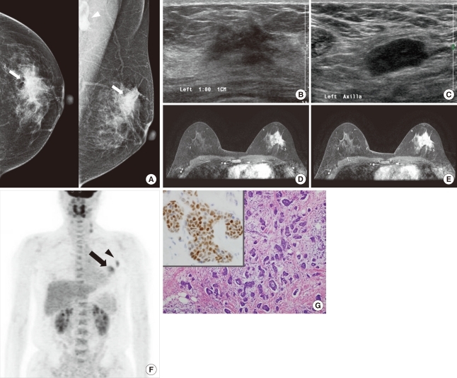
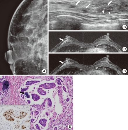
Most IMPCs presented as a high density irregular mass with a non-circumscribed margin associated with microcalcifications on mammography, as an irregular hypoechoic mass with a spiculated margin on ultrasound, and as irregular spiculated masses with washout patterns on MRI. PET-CT showed a high maximum standardized uptake value (SUVmax) (mean, 11.2). Axillary nodal metastases were identified in 65.5% of the patients. Immunohistochemical studies showed high positivities for estrogen receptor and c-erbB-2 (93.1% and 51.7µ, respectively).
Even though the imaging characteristics of IMPCs are not distinguishable from typical invasive ductal carcinomas, this tumor type frequently results in nodal metastases and high positivities for both estrogen receptor and c-erbB-2. The high SUVmax value that is apparent on PET-CT might be helpful in the diagnosis of IMPC.
本研究旨在评估包括乳腺浸润性微乳头状癌(IMPC)的影像学和组织病理学表现,包括免疫组化特征。
本研究纳入了 29 例经诊断为 IMPC 的患者。根据美国放射学院乳腺成像报告和数据系统词汇表,回顾性分析了乳腺 X 线摄影、超声和磁共振成像(MRI)的表现。(18)F-氟脱氧葡萄糖正电子发射断层扫描-计算机断层扫描(PET-CT)的结果也进行了评估。两位专门从事乳腺病理学的病理学家对手术标本的显微镜载玻片进行了共识审查。
大多数 IMPC 在乳腺 X 线摄影上表现为高密度不规则肿块,边界不清晰,伴有微钙化;在超声上表现为不规则低回声肿块,边界呈毛刺状;在 MRI 上表现为不规则的毛刺状肿块,伴有廓清模式。PET-CT 显示出高最大标准化摄取值(SUVmax)(平均值 11.2)。65.5%的患者存在腋窝淋巴结转移。免疫组织化学研究显示雌激素受体和 c-erbB-2 的阳性率较高(分别为 93.1%和 51.7µ)。
尽管 IMPC 的影像学特征与典型的浸润性导管癌无法区分,但这种肿瘤类型常导致淋巴结转移和雌激素受体及 c-erbB-2 的高阳性率。PET-CT 上明显的高 SUVmax 值可能有助于 IMPC 的诊断。